Friday, November 24, 2017

Pathophysiology of Cystic Fibrosis


Cystic fibrosis (CF
) is an inherited multisystem disorder of children and adults, characterized chiefly by obstruction and infection of airways and by maldigestion and its consequences.

CF is inherited as an autosomal recessive trait. The CF gene codes for a protein of 1,480 amino acids called the CF transmembrane regulator (CFTR).

Four long-standing observations are of fundamental pathophysiologic importance:
  1. failure to clear mucous secretions,
  2. a paucity of water in mucous secretions,
  3. an elevated salt content of sweat and other serous secretions, and
  4. chronic infection limited to the respiratory tract
In addition, there is a greater negative potential difference across the respiratory epithelia of CF patients than across the respiratory epithelia of control subjects. Aberrant electrical properties were also demonstrated for CF sweat gland duct epithelium. The membranes of CF epithelial cells are unable to secrete chloride ions in response to cyclic adenosine monophosphate (cAMP)–mediated signals and, at least in the respiratory tract, excessive amounts of sodium are absorbed through these membranes.

Cyclic AMP-stimulated chloride conductance is a function of CFTR itself; this function is absent in epithelial cells with many different mutations of the CFTR gene.

The postulated epithelial pathophysiology in airways involves an inability to secrete salt and secondarily to secrete water in the presence of excessive reabsorption of salt and water. The proposed outcome is insufficient water on the airway surface to hydrate secretions. Desiccated secretions become more viscous and elastic (rubbery) and are harder to clear by mucociliary and other mechanisms.These secretions are retained and obstruct airways, starting with those of the smallest caliber, the bronchioles. Airflow obstruction at the level of small airways is the earliest observable physiologic abnormality of the respiratory system.

Atrial Septal Defect


Introduction:

Atrial septal defects (ASDs) can occur in any portion of the atrial septum (secundum, primum, or sinus venosus), depending on which embryonic septal structure has failed to develop normally. Less commonly, the atrial septum may be nearly absent, with the creation of a functional single atrium. Isolated secundum ASDs account for ?7% of congenital heart defects.

The majority of cases of ASD are sporadic; autosomal dominant inheritance does occur as part of the Holt-Oram syndrome (hypoplastic or absent radii, 1st-degree heart block, ASD) or in families with secundum ASD and heart block.

Pathophysiology:

An isolated valve-incompetent patent foramen ovale (PFO) is a common echocardiographic finding during infancy. It is usually of no hemodynamic significance and is not considered an ASD; a PFO may play an important role if other structural heart defects are present. If another cardiac anomaly is causing increased right atrial pressure (pulmonary stenosis or atresia, tricuspid valve abnormalities, right ventricular dysfunction), venous blood may shunt across the PFO into the left atrium with resultant cyanosis. Because of the anatomic structure of the PFO, left-to-right shunting is unusual outside the immediate newborn period. In the presence of a large volume load or a hypertensive left atrium (secondary to mitral stenosis), the foramen ovale may be sufficiently dilated to result in a significant atrial left-to-right shunt. A valve-competent but probe-patent foramen ovale may be present in 15–30% of adults. An isolated PFO does not require surgical treatment, although it may be a risk for paradoxical (right to left) systemic embolization.

Neonatal Cold Injury


Neonatal cold injury usually occurs in abandoned infants or those in inadequately heated homes during damp cold spells when the outside temperature is in the freezing range .

Clinical Features:
The initial features are apathy, refusal of food, oliguria, and coldness to touch. The body temperature is usually between 29.5 and 35°C (85–95°F), and immobility, edema, and redness of the extremities, especially the hands, feet, and face, are observed. Bradycardia and apnea may also occur. The facial erythema frequently gives a false impression of health and delays recognition that the infant is ill. Local hardening over areas of edema may lead to confusion with scleredema. Rhinitis is common, as are hypoglycemia and acidosis. Hemorrhagic manifestations are frequent; massive pulmonary hemorrhage is a common finding at autopsy.

Treatment :

Consists of warming and paying scrupulous attention to recognizing and correcting hypotension and metabolic imbalances, particularly hypoglycemia.

Monday, November 20, 2017

Enterobius Vermicularis Infection in Children


ETIOLOGY.
The cause of enterobiasis, or pinworm infection, is Enterobius vermicularis,which is a small (1 cm in length), white, threadlike nematode, or roundworm, that typically inhabits the cecum, appendix, and adjacent areas of the ileum and ascending colon. Gravid females migrate at night to the perianal and perineal regions, where they deposit up to 15,000 eggs.Eggs embryonate within 6 hr and remain viable for 20 days.

MODE OF INFECTION.
Human infection occurs by the fecal-oral route typically by ingestion of embryonated eggs that are carried on fingernails, clothing, bedding, or house dust. After ingestion, the larvae mature to form adult worms in 36–53 days.

PATHOGENESIS.
Enterobius infection may cause symptoms by mechanical stimulation and irritation, allergic reactions, and migration of the worms to anatomic sites where they become pathogenic.

CLINICAL MANIFESTATIONS.
Pinworm infection is innocuous and rarely causes serious medical problems. The most common complaints include itching and restless sleep secondary to nocturnal perianal or perineal pruritus. The precise cause and incidence of pruritus are unknown. They may be related to the intensity of infection, psychologic profile of the infected individual and his or her family, or allergic reactions to the parasite.

Wednesday, November 15, 2017

Nail Separation


Onycholysis indicates separation of the nail plate from the distal nail bed.

Common causes are 
  • trauma, 
  • chronic exposure to moisture, 
  • hyperhidrosis, 
  • cosmetics, 
  • psoriasis, 
  • fungal infection (distal onycholysis), 
  • atopic or contact dermatitis, 
  • porphyria, 
  • drugs (bleomycin, vincristine, retinoid agents, indomethacin, chlorpromazine [Thorazine]), and 
  • drug-induced phototoxicity from tetracyclines or chloramphenicol.
Beau lines are transverse grooves in the nail plate that represent a temporary disruption of formation of the nail plate. The lines 1st appear a few weeks after the event that caused the disruption in nail growth. A single transverse ridge appears at the proximal nail fold in most 4–6 wk old infants and works its way distally as the nail grows; this line may reflect metabolic changes after delivery.

Clinical Manifestations of Congenital Hypopituitarism.


The child with hypopituitarism is usually of normal size and weight at birth although those with MPHD and genetic defects of the GH1 or GHR gene have birth lengths that average 1 SD below the mean. Children with severe defects in GH production or action are more than 4 SD below the mean by 1 yr of age. Those with less severe deficiencies grow at rates below the 25% for age and gradually diverge from normal height percentiles. Delayed closure of the epiphyses permits growth beyond the normal age when growth should be complete.

Infants with congenital defects of the pituitary or hypothalamus usually present with neonatal emergencies such as apnea, cyanosis, or severe hypoglycemia with or without seizures.

Microphallus in boys provides an additional diagnostic clue.

Deficiency of GH may be accompanied by hypoadrenalism and hypothyroidism.

Prolonged neonatal jaundice is common. It involves elevation of conjugated and unconjugated bilirubin and may be mistaken for neonatal hepatitis.

The head in the toddler is round, and the face is short and broad. The frontal bone is prominent, and the bridge of the nose is depressed and saddle-shaped. The nose is small, and the nasolabial folds are well developed. The eyes are somewhat bulging. The mandible and the chin are underdeveloped, and the teeth, which erupt late, are frequently crowded. The neck is short, and the larynx is small. The voice is high-pitched and remains high after puberty. The extremities are well proportioned, with small hands and feet. Weight for height is usually normal, but an excess of body fat and a deficiency of muscle mass contributes to a pudgy appearance.

Clinical Manifestations of Acquired Hypopituitarism


The child initially is normal; manifestations similar to those seen in idiopathic pituitary growth failure gradually appear and progress.

When complete or almost complete destruction of the pituitary gland occurs, signs of pituitary insufficiency are present.

Atrophy of the adrenal cortex, thyroid, and gonads results in loss of weight, asthenia, sensitivity to cold, mental torpor, and absence of sweating.

Sexual maturation fails to take place or regresses if already present. There may be atrophy of the gonads and genital tract with amenorrhea and loss of pubic and axillary hair.

There is a tendency to hypoglycemia. Growth slows dramatically. Diabetes insipidus may be present early but tends to improve spontaneously as the anterior pituitary is progressively destroyed.

Clinical Features of Cystic Fibrosis


The clinical features of cystic fibrosis are summarized below:

Respiratory Tract.Cough is the most constant symptom of pulmonary involvement. At first, the cough may be dry and hacking, but eventually it becomes loose and productive. In older patients, the cough is most prominent on arising in the morning or after activity. Expectorated mucus is usually purulent. Some patients remain asymptomatic for long periods or seem to have prolonged but intermittent acute respiratory infections. Others acquire a chronic cough in the 1st weeks of life, or they have pneumonia repeatedly.

Extensive bronchiolitis is attended by wheezing, which is a frequent symptom during the 1st years of life. As lung disease slowly progresses, exercise intolerance, shortness of breath, and failure to gain weight or grow are noted. Exacerbations of lung symptoms, presumably owing to more active airways infection, eventually require repeated hospitalizations for effective treatment.

Cor pulmonale, respiratory failure, and death eventually supervene unless lung transplantation is accomplished. Colonization with Burkholderia cepacia and other multidrug-resistant organisms may be associated with particularly rapid pulmonary deterioration and death.

Early physical findings include increased anteroposterior diameter of the chest, generalized hyperresonance, scattered or localized coarse crackles, and digital clubbing. Expiratory wheezes may be heard, especially in young children.

Even though the paranasal sinuses are virtually always opacified radiographically, acute sinusitis is infrequent. Nasal obstruction and rhinorrhea are common, caused by inflamed, swollen mucous membranes or, in some cases, nasal polyposis. Nasal polyps are most troublesome between 5 and 20 yr of age.

Sunday, November 12, 2017

Endocardial Cushion Defect - Pathophysiology, Clinical findings And Management


Pathophysiology

– When both ASDs and VSDs occur, which are contiguous, and the atrioventricular valves are abnormal
Left-to-right shunt at both atrial and ventricular levels; some right-to-left shunting with desaturation (mild, intermittent cyanosis)
Atrioventricular valve insufficiency –+ increase volume load on one or both ventricles; early heart failure, infections, minimal cyanosis, hepatomegaly, and failure to thrive.

Physical examination

Heart failure early in infancy (hepatomegaly, failure to thrive)
Eisenmenger physiology occurs earlier
– Moderate-to-severe increase in heart size with hyperdynamic precordium (precordial bulge and lift)
– Loud S1 widely fixed split S2 (like an isolated ASD)
Pulmonary systolic ejection murmur, low-pitched diastolic rumble at left sternal border and apex; may also have mitral insufficiency (apical harsh holosystolic murmur radiating to left axilla).

Clinical Features of Diseases Caused by Shigella


Bacillary dysentery
is clinically similar regardless of infecting serotype; however, there are some clinical differences, particularly relating to the greater severity and risk for complications with S. dysenteriaeserotype 1 infection.

Incubation Period:
Ingestion of shigellae is followed by an incubation period of 12 hr to several days before symptoms ensue.

Clinical Manifestations:
Severe abdominal pain, high fever, emesis, anorexia, generalized toxicity, urgency, and painful defecation characteristically occur. Physical examination at this point may show abdominal distention and tenderness, hyperactive bowel sounds, and a tender rectum on digital examination.

The diarrhea may be watery and of large volume initially, evolving into frequent small-volume, bloody mucoid stools; most children (>50%) never progress to the stage of bloody diarrhea, whereas in others the 1st stools are bloody. Significant dehydration related to the fluid and electrolyte losses in both feces and emesis can occur. Untreated diarrhea may last 1–2 wk; only about 10% of patients have diarrhea persisting for more than 10 days.

Neurologic findings are among the most common extraintestinal manifestations of bacillary dysentery, occurring in as many as 40% of hospitalized infected children. Enteroinvasive E. coli can cause similar neurologic toxicity. Convulsions, headache, lethargy, confusion, nuchal rigidity, or hallucinations may be present before or after the onset of diarrhea. The cause of these neurologic findings is not understood.

Clinical Features of Infectious Mononucleosis


Incubation Period:

The incubation period of infectious mononucleosis in adolescents is 30–50 days. In children, it may be shorter. The majority of cases of primary EBV infection in infants and young children are clinically silent. In older patients, the onset of illness is usually insidious and vague.

Prodrome of Illness:
Patients may complain of malaise, fatigue, acute or prolonged (>1 wk) fever, headache, sore throat, nausea, abdominal pain, and myalgia. This prodromal period may last 1–2 wk. The complaints of sore throat and fever gradually increase until patients seek medical care. Splenic enlargement may be rapid enough to cause left upper quadrant abdominal discomfort and tenderness, which may be the presenting complaint.

Physical Examination:

The physical examination is characterized by generalized lymphadenopathy (90% of cases), splenomegaly (50% of cases), and hepatomegaly (10% of cases).

Lymphadenopathy occurs most commonly in the anterior and posterior cervical nodes and the submandibular lymph nodes and less commonly in the axillary and inguinal lymph nodes. Epitrochlear lymphadenopathy is particularly suggestive of infectious mononucleosis.

Symptomatic hepatitis or jaundice is uncommon, but elevated liver enzymes are common.

Splenomegaly to 2–3 cm below the costal margin is typical; massive enlargement is uncommon.

Diagnosis of Infectious Mononucleosis


The diagnosis of infectious mononucleosis implies primary EBV infection.

A presumptive diagnosis may be made by the presence of typical clinical symptoms with atypical lymphocytosis in the peripheral blood. The diagnosis is usually confirmed by serologic testing, either for heterophile antibody or specific EBV antibodies.

Culture of EBV is tedious and requires 4–6 wk. The culture method is the transformation assay, which is performed by co-cultivating oropharyngeal or genital secretions, peripheral blood (10–30 mL), or tumor with human umbilical cord lymphocytes. The cultures are observed for 6 wk for signs of cell transformation: proliferation and rapid growth, mitotic figures, large vacuoles, granular morphology, and cell aggregation. EBV immortalizes the umbilical cord cells, resulting in cell lines that can be maintained in perpetuity that harbor EBV isolated from the patient.

In >90% of cases there is leukocytosis of 10,000–20,000 cells/mm3, of which at least ? are lymphocytes; atypical lymphocytes usually account for 20–40% of the total number. The atypical cells are mature T lymphocytes that have been antigenically activated. Compared with regular lymphocytes microscopically, atypical lymphocytes are larger overall, with larger, eccentrically placed indented and folded nuclei with a lower nuclear-to-cytoplasm ratio. Although atypical lymphocytosis may be seen with many of the infections usually causing lymphocytosis, the highest degree of atypical lymphocytes is classically seen with EBV infection.

Heterophile antibodies agglutinate cells from species different from those in the source serum. The transient heterophile antibodies seen in infectious mononucleosis, also known as Paul-Bunnell antibodies, are IgM antibodies detected by the Paul-Bunnell-Davidsohn test for sheep red cell agglutination. The heterophile antibodies of infectious mononucleosis agglutinate sheep or, for greater sensitivity, horse red cells but not guinea pig kidney cells.

Scurvy in Children


Very low intake of vitamin C over time may lead to the deficiency disease scurvy.

Age of Onset:
In infants and young children, the usual age of onset of clinical manifestations of scurvy is 6–24 months.

Early Symptoms:
The early symptoms are rather general and include low-grade fever, irritability, tachypnea, digestive disturbances, loss of appetite, and generalized tenderness, particularly in the legs, which is noticeable when the diaper is changed. The pain results in pseudoparalysis, with the hips and knees semi-flexed and the feet rotated outward.

Other Clinical Features:

Edematous swelling along the shafts of the legs may be present; in some cases, there is subperiosteal hemorrhage at the end of the femur .

A “rosary” at the costochondral junctions and depression of the sternum are other typical features.

Changes in the gums are most noticeable after teeth have erupted and are manifested as bluish purple, spongy swellings of the mucous membrane, especially over the upper incisors.

Anemia, which is seen primarily in infants and young children, may be related to impaired ability to use iron or folate.

Other clinical manifestations seen in infants as well as in older children and adolescents include swollen joints, purpura and ecchymoses, poor wound and fracture healing, petechiae, perifollicular hemorrhages , hyperkeratosis of hair follicles, arthralgia, and muscle weakness. Endochondral bone formation may not proceed because osteoblasts cannot form osteoid. Bony trabeculae that have been formed become brittle and fracture easily. Irritability and other psychologic manifestations are likely due to impaired neurotransmitter metabolism.

Brain Abscess In Children


Brain abscesses can occur in children of any age but are most common in children between 4 and 8 yr and neonates.

CAUSES.
The causes of brain abscess include embolization due to congenital heart disease with right-to-left shunts (especially tetralogy of Fallot), meningitis, chronic otitis media and mastoiditis, sinusitis, soft tissue infection of the face or scalp, orbital cellulitis, dental infections, penetrating head injuries, immunodeficiency states, and infection of ventriculoperitoneal shunts.

PATHOLOGY.
Cerebral abscesses are evenly distributed between the two hemispheres, and aapx. 80% of cases are divided equally between the frontal, parietal, and temporal lobes.. An abscess in the frontal lobe is often caused by extension from sinusitis or orbital cellulitis, whereas abscesses located in the temporal lobe or cerebellum are frequently associated with chronic otitis media and mastoiditis.

ETIOLOGY.
The responsible bacteria include streptococci (S. milleri, S. pyogenes group A or B, S. pneumoniae, S. faecalis), anaerobic organisms (gram-positive cocci, Bacteroides spp., Fusobacterium spp., Prevotella spp., Actinomyces spp.), and gram-negative aerobic bacilli (Haemophilus aphrophilus, H. parainfluenzae, H. influenzae, Enterobacter, E. coli, Proteus spp.). Citrobacter is most common in neonates. One organism is cultured in the majority of abscesses (70%), two in 20%, and three or more in 10% of cases. Abscesses associated with mucosal infections (sinusitis) frequently have anaerobic bacteria.

Monday, November 6, 2017

Treatment of Iron Deficiency Anemia


The regular response of iron-deficiency anemia to adequate amounts of iron is an important diagnostic and therapeutic feature.

Oral Iron:
Oral administration of simple ferrous salts (e.g., sulfate, gluconate, fumarate) provides inexpensive and satisfactory therapy. No evidence shows that addition of any trace metal, vitamin, or other hematinic substance significantly increases the response to simple ferrous salts. One problem encountered with administration of oral iron to young children is that liquid FeSO4nhas an unpleasant taste, but sometimes the taste can be camouflaged by mixing with flavored syrup. Other, better-tasting preparations are available over the counter, but these are much more expensive than simple liquid FeSO4. Aside from the unpleasant taste, intolerance to oral iron is uncommon in young children, although older children and adolescents sometimes have gastrointestinal complaints. Problems with constipation can be minimized by increasing water and fiber intake. For some children, abdominal discomfort can be minimized by administering iron with food, recognizing that this may decrease iron absorption to some extent.

The therapeutic dose should be calculated in terms of elemental iron; ferrous sulfate is 20% elemental iron by weight. A daily total dose of 4–6 mg/kg of elemental iron in 3 divided doses provides an optimal amount of iron for the stimulated bone marrow to use.

Myocarditis


Myocarditis
refers to inflammation, necrosis, or myocytolysis and may be caused by many infectious, connective tissue, granulomatous, toxic, or idiopathic processes affecting the myocardium with or without associated systemic manifestations of the disease process or involvement of the endocardium or pericardium.

Etiology:
The most common causative agents in children are adenovirus, coxsackievirus B, and other enteroviruses, although most known viral agents have been implicated.

Pathophysiology:
Acute viral myocarditis may produce a fulminant inflammatory process characterized by cellular infiltrates, cell degeneration and necrosis, and subsequent fibrosis. Viral myocarditis may also become a chronic process with persistence of viral RNA or DNA (but not infectious virus particles) in the myocardium.In addition, some viral proteins may share antigenic epitopes with host cells, which results in autoimmune damage to the antigenically related myocyte.

The net final result of chronic viral-associated inflammation is often dilated cardiomyopathy.

Clinical Manifestations:

Signs and symptoms depend on the patient’s age and the acute or chronic nature of the infection. A neonate may initially have fever, severe heart failure, respiratory distress, cyanosis, distant heart sounds, weak pulses, tachycardia out of proportion to the fever, mitral insufficiency caused by dilatation of the valve annulus, a gallop rhythm, acidosis, and shock. Evidence of viral hepatitis, aseptic meningitis, and an associated rash may be present. In the most fulminant form, death may occur within 1–7 days of the onset of symptoms.

Treatment of Pneumothorax


Therapy varies with the extent of the collapse and the nature and severity of the underlying disease.

A tension pneumothorax may emergently require drainage by needle thoracostomy. A small or even moderate-sized pneumothorax in an otherwise normal child may resolve without specific treatment, usually within about 1 wk. A small (<5%) pneumothorax complicating asthma may also resolve spontaneously.

Administering 100% oxygen may hasten resolution. Patients with chronic hypoxemia should be monitored closely during the administration of supplemental oxygen. Pleural pain deserves analgesic treatment. If there is >5% collapse or if the pneumothorax is recurrent or under tension, chest tube drainage is necessary.

Pneumothoraces complicating Cystic fibrosis frequently recur, and definitive treatment may be justified with the 1st episode, even with <5% collapse.

Closed thoracotomy (simple insertion of a chest tube) and drainage of the trapped air through a catheter, the external opening of which is kept in a dependent position under water, is adequate to re-expand the lung in most patients; pigtail catheters are frequently used.

When there have been previous pneumothoraces, it may be indicated to induce the formation of strong adhesions between the lung and chest wall by a sclerosing procedure to prevent recurrence. This can be carried out by the introduction of doxycycline or talc into the pleural space (chemical pleurodesis).

Clinical Manifestations of Osteogenesis Imperfecta


Osteogenesis imperfecta
has the triad of fragile bones, blue sclerae, and early deafness. OI was once divided into “congenita,” the forms detectable at birth, and “tarda,” the forms detectable later in childhood; this did not account for the variability of OI.

The Sillence classification divides OI into four types based on clinical and radiographic criteria. Additional types have been proposed based on histologic distinctions.

Osteogenesis Imperfecta Type I (Mild):
This form is sufficiently mild that it is often found in large pedigrees. Many type I families have blue sclerae, recurrent fractures in childhood, and presenile hearing loss (30–60%). Both types I and IV are divided into A and B subtypes, depending on the absence (A) or presence (B) of dentinogenesis imperfecta. Other possible connective tissue abnormalities include easy bruising, joint laxity, and mild short stature compared with family members. Fractures result from mild to moderate trauma and decrease after puberty.

Osteogenesis Imperfecta Type II (Perinatal Lethal):

These infants may be stillborn or die in the 1st yr of life. Birthweight and length are small for gestational age. There is extreme fragility of the skeleton and other connective tissues. There are multiple intrauterine fractures of long bones, which have a crumpled appearance on radiographs. There are striking micromelia and bowing of extremities; the legs are held abducted at right angles to the body in the “frog-leg position.” Multiple rib fractures create a beaded appearance and the small thorax contributes to respiratory insufficiency. The skull is large for body size with enlarged anterior and posterior fontanelles. Sclerae are dark blue-gray

Splenomegaly


A soft, thin spleen may be palpable in 15% of neonates, 10% of normal children, and 5% of adolescents. In most individuals, the spleen must be 2–3 times its normal size before it is palpable.

Examination:
The spleen is best examined in a supine patient by palpating across the abdomen toward the left costal margin from below as the patient inspires deeply. An enlarged spleen might descend into the pelvis; when splenomegaly is suspected, the abdominal examination should begin at a lower starting point. Superficial abdominal venous distention may be present when splenomegaly is a result of portal hypertension.

Radiologic detection or confirmation of splenic enlargement is done with ultrasonography, CT, or technetium-99 sulfur colloid scan. The latter also assesses splenic function.

Pseudosplenomegaly
.
Abnormally enlarged mesenteric connections may produce a wandering or ptotic spleen. An enlarged left lobe of the liver, a left upper quadrant mass, or a splenic hematoma may be mistaken for splenomegaly. Splenic cysts may contribute to splenomegaly or mimic it; these may be congenital (epidermoid) or acquired (pseudocyst) after trauma or infarction. Cysts are usually asymptomatic and are found on radiologic evaluation. Splenosis after splenic rupture or an accessory spleen (present in 10% of normal individuals) may also mimic splenomegaly; most are not palpable.

Giardiasis in Children


Giardia lamblia
is a flagellated protozoan that infects the duodenum and small intestine. Infection results in clinical manifestations that range from asymptomatic colonization to acute or chronic diarrhea and malabsorption. Infection is more prevalent in children than in adults.

Life Cycle:

The life cycle of G. lamblia (also known as Giardia intestinalis or Giardia duodenalis) is composed of 2 stages: trophozoites and cysts.

Giardia infects humans after ingestion of as few as 10–100 cysts. Ingested cysts, which measure 8–10 mm in diameter, each produce 2 trophozoites in the duodenum. After excystation, trophozoites colonize the lumen of the duodenum and proximal jejunum, where they attach to the brush border of the intestinal epithelial cells and multiply by binary fission. The body of the trophozoite is teardrop shaped, measuring 10–20 mm in length and 5–15 mm in width. Giardia trophozoites contain 2 oval nuclei anteriorly, a large ventral disk, a curved median body posteriorly, and 4 pairs of flagella. As detached trophozoites pass down the intestinal tract, they encyst to form oval cysts that contain 4 nuclei.

Cysts are passed in stools of infected individuals and may remain viable in water for as long as 2 months. Their viability often is not affected by the usual concentrations of chlorine used to purify water for drinking.

Clinical Manifestations:

The incubation period of Giardia infection usually is 1–2 wk but may be longer. A broad spectrum of clinical manifestations occurs, depending on the interaction between G. lamblia and the host. Children who are exposed to G. lamblia may experience asymptomatic excretion of the organism, acute infectious diarrhea, or chronic diarrhea with persistent gastrointestinal tract signs and symptoms, including failure to thrive and abdominal pain or cramping. Most infections in both children and adults are asymptomatic.

Alopecia in Children


True alopecia
is rarely congenital; it is more often related to an inflammatory dermatosis, mechanical factors, drug ingestion, infection, endocrinopathy, nutritional disturbance, or disturbance of the hair cycle. Any inflammatory condition of the scalp, such as atopic dermatitis or seborrheic dermatitis, if severe enough, may result in partial alopecia; hair growth returns to normal if the underlying condition is treated successfully, unless the hair follicle has been permanently damaged.

TRACTION ALOPECIA.
Traction alopecia is due to trauma to hair follicles from tight braids or ponytails, headbands, rubber bands, curlers, or rollers . Children and parents must be encouraged to avoid devices that cause trauma to the hair and, if necessary, to alter the hairstyle. Otherwise, scarring of hair follicles may occur.

HAIR PULLING.
Hair pulling in childhood is usually an acute reactional process related to emotional stress. It may also be seen in trichotillomania and as part of more severe psychiatric disorders.

TRICHOTILLOMANIA.
Compulsive pulling, twisting, and breaking of hair produces irregular areas of incomplete hair loss, most often on the crown and in the occipital and parietal areas of the scalp. Occasionally, eyebrows, eyelashes, and body hair are traumatized. Some plaques of alopecia may have a linear outline. The hairs remaining within the areas of loss are of various lengths and are typically blunt tipped because of breakage. The scalp usually appears normal, although hemorrhage, crusting and chronic folliculitis may also occur.

Sunday, November 5, 2017

Laboratory Finding In Hereditary Spherocytosis


The different laboratory findings observed in patients with hereditary spherocytosis is summarized as follows.

Evidence of hemolysis includes reticulocytosis and indirect hyperbilirubinemia.

Hb Level:
The hemoglobin level usually is 6–10 g/dL, but it can be in the normal range.

Retic Count:
The reticulocyte percentage often is increased to 6–20%, with a mean of approximately 10%.

Absolute Values:

The mean corpuscular volume is normal, although the mean corpuscular hemoglobin concentration often is increased (36–38 g/dL RBCs).

Blood Film:

The RBCs on the blood film vary in size and include polychromatophilic reticulocytes and spherocytes. The spherocytes are smaller in diameter and appear hyperchromic on the blood film as a result of the high hemoglobin concentration. The central pallor is less conspicuous than in normal cells. Spherocytes may be the predominant cell or may be relatively sparse, depending on the severity of the disease, but they usually account for >15–20% of the cells when hemolytic anemia is present.

Bone marrow:
Erythroid hyperplasia is evident in the marrow aspirate or biopsy. Marrow expansion may be evident on routine roentgenographic examination.

Other Features :
Other evidence of hemolysis may include decreased haptoglobin and the presence of gallstones on ultrasonography.

Pulmonary Edema


Pulmonary edema
is an excessive accumulation of fluid in the interstitium and air spaces of the lung, resulting in oxygen desaturation and respiratory distress. It is a common problem in the acutely ill child and a sequela of several different pathologic processes.

Pathophysiology
Although pulmonary edema is traditionally separated into two categories according to cause (cardiogenic and noncardiogenic), the end result of both processes is a net fluid accumulation within the interstitial and alveolar spaces. Noncardiogenic pulmonary edema, in its most severe state, is also known as acute respiratory distress syndrome (ARDS).

The pressure and osmolarity on either side of a pulmonary capillary and capillary permeability are the physical factors that determine fluid movement through the capillary wall. Baseline conditions lead to a net filtration of fluid from the intravascular space into the interstitium. This “extra” interstitial fluid is usually rapidly reabsorbed by pulmonary lymphatics. Conditions that lead to altered capillary permeability, increased pulmonary capillary pressure, or decreased intravascular oncotic pressure increase the net flow of fluid out of the capillary and thereby lead to water accumulation in the lung.

Nasal Polyps in Children


Nasal polyps are benign pedunculated tumors formed from edematous, usually chronically inflamed nasal mucosa. They commonly arise from the ethmoidal sinus and present in the middle meatus. Occasionally, they appear within the maxillary antrum and can extend to the nasopharynx.

Incidence:
Whereas antrochoanal polyps represent only 4–6% of all nasal polyps in the general population, they represent about 33% of polyps in the pediatric population.

Etiology:
Cystic fibrosis is the most common childhood cause of nasal polyposis and should be suspected in any child <12 yr old with nasal polyps, even in the absence of typical respiratory and digestive symptoms; as many as 30% of children with cystic fibrosis acquire nasal polyps.

Nasal polyposis is also associated with chronic sinusitis and allergic rhinitis.

In the uncommon Samter triad, nasal polyps are associated with aspirin sensitivity and asthma.

Clinical Manifestations:
Obstruction of nasal passages is prominent, with associated hyponasal speech and mouth breathing. Profuse unilateral mucoid or mucopurulent rhinorrhea may also be present. An examination of the nasal passages shows glistening, gray, grapelike masses squeezed between the nasal turbinates and the septum.

Diagnosis:Examination of the external nose and rhinoscopy is performed. Ethmoidal polyps can be readily distinguished from the well-vascularized turbinate tissue, which is pink or red; antrochoanal polyps may have a more fleshy appearance. Antrochoanal polyps may prolapse into the nasopharynx; flexible nasopharyngoscopy can assist in making this diagnosis.

CT scan of the midface is key to diagnosis and planning for surgical treatment.

Friday, November 3, 2017

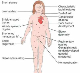
Turner’s Syndrome


Genetics

– Generally sporadic; no older maternal age seen
– Paternal chromosome more likely to be missing

Findings
– Small-stature female
-Gonadal dysgenesis
– Tendency to become obese
– Average IQ =90
– Decreased motor skills, incoordination
– Congenital lymphedema, residual puffiness over dorsum of fingers and toes
– Broad chest, wide-spaced nipples
– Low posterior hairline
– Webbed posterior neck
– Cubitus valgus (elbow)
– Horseshoe kidney, double renal pelvis
– Cardiac:
o Bicuspid aortic valve
o Coarctation
o Aortic stenosis, mitral valve prolapse
o Hypertension common, even without cardiac or renal disease.

Allergic Rhinitis


Generally established by 6 years of age.

Increased risk
– early introduction of formula (versus breast milk) or solids, mother smoking before child is 1 year old, heavy exposure to indoor allergens.
-Most perennial or mixed.
-Increased symptoms with greater exposure.
.-Diagnosis suggested by typical symptoms in absence of URI or structural abnormality (nasal congestion/pruritus, worse at night with snoring, mouth-breathing; watery, itchy eyes; postnasal drip with cough; possible wheezing; headache)
Specific behaviors
– Allergic salute (rhinorrhea and nasal pruritus) ~ nasal crease
– Vigorous grinding of eyes with thumb and side of fist.

• History of symptoms

– Timing and duration (seasonal versus perennial)
– Exposures/settings in which symptoms occur
– Family history of allergic disease (atopy, asthma)
– Ask about perennial versus seasonal
– Seasonal allergies-usually need several seasons of exposure
– Food allergies more common (nuts, seafood) in young children (then skin, gastrointestinal, and, less often, respiratory)

Different Clinical Features in Children with SLE


Children with lupus present with diverse and often severe manifestations. Children most frequently present with fever, fatigue, hematologic abnormalities, arthralgia or arthritis, rash, and renal disease. Symptoms may be intermittent or persistent.

Cutaneous manifestations are frequently present. The characteristic malar or butterfly rash involves the cheeks and nasal bridge and varies from an erythematous blush to thickened epidermis to scaly patches . Rashes may be photosensitive and extend to all sun-exposed areas.

Mucous membrane changes ranging from vasculitic erythema to ulcers occur, particularly on palatal and nasal mucosa.

Musculoskeletal findings include arthralgia, arthritis, tendinitis, and myositis. Deforming arthritis is unusual, although hand arthritis can lead to ligament damage and severely lax joints.

Serositis can affect pleural, pericardial, and peritoneal surfaces. Hepatosplenomegaly and lymphadenopathy are often found. Other gastrointestinal manifestations, most often resulting from vasculitis, include pain, diarrhea, melena, infarction, inflammatory bowel disease, and hepatitis.

Treatment for Tetanus in Children


Management of tetanus requires eradication of C. tetani and the wound environment conducive to its anaerobic multiplication, neutralization of all accessible tetanus toxin, control of seizures and respiration, palliation and provision of meticulous supportive care, and, finally, prevention of recurrences.

Surgical wound excision and debridement are often needed to remove the foreign body or devitalized tissue that created anaerobic growth conditions.

Tetanus toxin cannot be neutralized by TIG after it has begun its axonal ascent to the spinal cord. TIG should be given as soon as possible in order to neutralize toxin that diffuses from the wound into the circulation before the toxin can bind at distant muscle groups. The optimal dose of TIG has not been determined. A single intramuscular injection of 500 U of TIG is sufficient to neutralize systemic tetanus toxin, but total doses as high as 3,000–6,000 U are also recommended. Infiltration of TIG into the wound is now considered unnecessary.

If TIG is unavailable, use of human intravenous immunoglobulin (IVIG), may be necessary. IVIG contains 4–90 U/mL of TIG; the optimal dosage of IVIG for treating tetanus is not known and it is not approved for this indication.

Another alternative is equine- or bovine-derived tetanus antitoxin (TAT). The usual dose of TAT is 50,000–100,000 U, with half given intramuscularly and half intravenously, but as little as 10,000 U may be sufficient. TAT is not available in the USA. Approximately 15% of patients given the usual dose of TAT experience serum sickness. When using TAT, it is essential to check for possible sensitivity to horse serum and desensitization may be needed. The human-derived immunoglobulins are much preferred because of their longer half-life (30 days) and the virtual absence of allergic and serum sickness adverse effects.

Vomiting in a Newborn Baby


Vomiting or, more often, regurgitation is a relatively frequent symptom during the neonatal period. In the 1st few hours after birth, infants may vomit mucus, occasionally blood streaked. This vomiting rarely persists after the 1st few feedings; it may be due to irritation of the gastric mucosa by material swallowed during delivery. If vomiting is protracted, gastric lavage with physiologic saline solution may relieve it.

When vomiting occurs shortly after birth and is persistent, the possibilities of intestinal obstruction, metabolic disorders, and increased intracranial pressure must be considered. A history of maternal polyhydramnios suggests upper gastrointestinal (esophageal, duodenal, ileal) atresia. Bile-stained emesis suggests intestinal obstruction beyond the duodenum, but may also be idiopathic.

Obstructive lesions of the digestive tract are the most frequent gastrointestinal anomalies.

Vomiting from esophageal obstruction occurs with the 1st feeding. The diagnosis of esophageal atresia can be suspected if unusual drooling from the mouth is observed and if resistance is encountered during an attempt to pass a catheter into the stomach. The diagnosis should be made before the infant has trouble with oral feedings and develops aspiration pneumonia.

Infantile achalasia (cardiospasm), a rare cause of vomiting in newborn infants, is demonstrable roentgenographically by obstruction at the cardiac end of the esophagus without organic stenosis. Regurgitation of feedings because of continuous relaxation of the esophageal-gastric sphincter, or chalasia, is a cause of vomiting. Keeping the infant in a semi-upright position, thickening the feeding, or administering prokinetic drugs can control it.

Vomiting because of obstruction of the small intestine usually begins on the 1st day of life and is frequent, persistent, usually non-projectile, copious, and, unless the obstruction is above the ampulla of Vater, bile stained; it is associated with abdominal distention, visible deep peristaltic waves, and reduced or absent bowel movements.

Clinical Features of Hepatitis A in Children


HAV is responsible for acute hepatitis only. Often, this is an anicteric illness, with clinical symptoms indistinguishable from other forms of viral gastroenteritis, particularly in young children.

The illness is much more likely to be symptomatic in older adolescents or adults, in patients with underlying liver disorders, and in those who are immunocompromised. It is characteristically an acute febrile illness with an abrupt onset of anorexia, nausea, malaise, vomiting, and jaundice.

Duration of Illness: The typical duration of illness is 7–14 days

Other organ systems can be affected during acute HAV infection. Regional lymph nodes and the spleen may be enlarged. The bone marrow may be moderately hypoplastic, and aplastic anemia has been reported. Small intestinal tissue may show changes in villous structure, and ulceration of the gastrointestinal tract can occur, especially in fatal cases. Acute pancreatitis and myocarditis have been reported, though rarely, and nephritis, arthritis, vasculitis, and cryoglobulinemia can result from circulating immune complexes.

Complications: Although most patients achieve full recovery, two distinct complications can occur:

Laboratory Findings in Primary Aldosteronism


Primary aldosteronism encompasses disorders caused by excessive aldosterone secretion independent of the renin-angiotensin system. These disorders are characterized by hypertension, hypokalemia,and suppression of the renin-angiotensin system
  • Hypokalemia occurs frequently.
  • Serum pH and the carbon dioxide and sodium concentrations may be elevated and 
  • the serum chloride and magnesium levels decreased. 
  • Serum levels of calcium are normal, even in children who manifest tetany. 
  • The urine is neutral or alkaline, and urinary potassium excretion is high. 
  • Plasma levels of aldosterone may be normal or elevated. 
  • Aldosterone concentrations in 24-hr urine collections are always increased. 
  • Plasma levels of renin are persistently low.
The diagnostic test of choice for primary aldosteronism is controversial. Both renin and aldosterone levels may vary by time of day, posture, and sodium intake, making it difficult to establish consistent reference ranges. It is desirable to establish a consistent sampling protocol, for example, mid-morning after the patient has been sitting for 15 min. If possible, antihypertensive drugs or other medications that can affect aldosterone or renin secretion should be avoided for several weeks prior to testing,

Fetal Alcohol Syndrome


High levels of alcohol ingestion during pregnancy can be damaging to embryonic and fetal development.

Incidence:

A specific pattern of malformation identified as fetal alcohol syndrome has been documented, and major and minor components of the syndrome are expressed in 1–2 infants/1,000 live births.

Both moderate and high levels of alcohol intake during early pregnancy may result in alterations in growth and morphogenesis of the fetus; the greater the intake, the more severe the signs. Infants born to heavy drinkers have twice the risk of abnormality as those born to moderate drinkers; 32% of infants born to heavy drinkers had congenital anomalies as compared with 9% in the abstinent and 14% in the moderate group. Additional maternal risk factors associated with fetal alcohol syndrome are advanced maternal age, low socioeconomic status, poor psychologic indicators, and binge drinking.

Clinical Features:

Characteristics of fetal alcohol syndrome include

(1) prenatal onset and persistence of growth deficiency for length, weight, and head circumference;

(2) facial abnormalities, including short palpebral fissures, epicanthal folds, maxillary hypoplasia, micrognathia, smooth philtrum, and a thin, smooth upper lip ;

(3) cardiac defects, primarily septal defects;

(4) minor joint and limb abnormalities, including some restriction of movement and altered palmar crease patterns; and 

(5) delayed development and mental deficiency varying from borderline to severe.

Wednesday, November 1, 2017

Diagnosis of Sickle Cell Disease


Virtually all states in the U.S. have instituted mandatory newborn screening program for sickle cell disease. Such programs identify newborns with the disease, provide prompt diagnosis and anticipatory guidance for the parents, and make possible the initiation of treatment with penicillin before 4 mo of age.

Newborn Screening:

The most commonly used procedures for newborn diagnosis include thin-layer/isoelectric focusing and high-performance liquid chromatography. A 2-step system is recommended, in which all initially abnormal screens are retested during the 1st clinical visit and again after 6 mo of age to determine the final hemoglobin phenotype.

A complete blood cell count as well as hemoglobin analysis is recommended on both parents to confirm the diagnosis and provide an opportunity for genetic counseling.

In affected patients, the red blood cell morphology after 3–6 mo of life is helpful for sickle cell disease and other hemoglobinopathies.

Newborn screening programs 1st report the hemoglobin with the greatest quantity, followed by the other hemoglobins in decreasing quantity. In newborns with a hemoglobin analysis consistent with a diagnosis of sickle cell disease, the FS pattern is supportive of Hb SS, Hb hereditary persistent fetal hemoglobin (Hb S/?0). The FSA pattern is supportive of the diagnosis of Hb S/?+. The diagnosis of Hb S/?+is confirmed if at least 50% of hemoglobin is Hb S, Hb A is present, and an elevated amount of Hb A2is present (typically >3.5%).

Candida Infection in Newborn


Candida is a common cause of oral mucous membrane infections (thrush) and perineal skin infections (candidal diaper dermatitis) in newborn infants. Disseminated candidiasis and candidemia have become a frequent problem in neonatal intensive care units.

Epidemiology.
C. albicans is commonly part of the gastrointestinal and vaginal flora of adults. Pregnancy increases the rate of maternal vaginal colonization from <20% to >30%. Maternal colonization rates at time of delivery correlate with the colonization rates of the newborns. Approximately 10% of full-term infants become colonized in the gastrointestinal and respiratory tract in the 1st 5 days of life. Neonatal risk factors for invasive candidiasis include very low birthweight status, broad-spectrum antibiotic administration, abdominal surgery, prolonged ventilatory support, prolonged intravenous catheterization, and parenteral alimentation.

Pathogenesis
The inability of the newborn infant to localize, control, and eradicate Candida infections is related to the relative impairment of specific and nonspecific host defense mechanisms. Overgrowth of Candida on mucocutaneous surfaces and colonization of intravenous catheters favor entry and penetration, with development of clinical infection directly related to extent of colonization. Hematogenous spread may lead to vasculitis and miliary nodules in multiple organs, including the skin, liver, spleen, lungs, kidneys, gastrointestinal tract, heart, eyes, and meninges.

Introduction To Abetalipoprotienemia


This rare autosomal recessive disorder of lipoprotein metabolism is associated with severe fat malabsorption from birth.

Clinical Features:

Children fail to thrive during the 1st year of life, and their stools are pale, foul smelling, and bulky. The abdomen is distended, and deep tendon reflexes are absent as a result of peripheral neuropathy, which is secondary to vitamin E deficiency. Intellectual development tends to be slow. After 10 yr of age, intestinal symptoms are less severe, ataxia develops, and there is a loss of position and vibration sensation with the onset of intention tremors. These last symptoms reflect involvement of the posterior columns, cerebellum, and basal ganglia. In adolescence, atypical retinitis pigmentosa develops.

Diagnosis:

Diagnosis rests on finding acanthocytes in the peripheral blood and extremely low plasma levels of cholesterol (<50 mg/dL); triglycerides are very low (<20 mg/dL). Chylomicrons and very low density lipoproteins are not detectable, and the low-density lipoprotein (LDL) fraction is virtually absent from the circulation; marked triglyceride accumulation in villus enterocytes occurs in the fasting duodenal mucosa. Steatorrhea occurs in younger patients, but other processes of assimilation are intact. Rickets may be an unusual initial manifestation of abetalipoproteinemia and hypobetalipoproteinemia. Rickets is caused by steatorrhea-induced calcium losses. Patients have mutations of the microsomal triglyceride transfer protein (MTP) gene, resulting in absence of MTP function in the small bowel. This protein is required for normal assembly and secretion of very low density lipoproteins and chylomicrons.

Clinical Manifestations of Bronchiolitis


Acute bronchiolitis
is usually preceded by exposure to an older contact with a minor respiratory syndrome within the previous wk.

Intial Symptoms: The infant 1st develops a mild upper respiratory tract infection with sneezing and clear rhinorrhea.

Fever: This may be accompanied by diminished appetite and fever of 38.5–39°C (101–102°F), although the temperature may range from subnormal to markedly elevated.

Respiratory symptoms: Gradually, respiratory distress ensues, with paroxysmal wheezy cough, dyspnea, and irritability. The infant is often tachypneic, which may interfere with feeding. The child does not usually have other systemic complaints, such as diarrhea or vomiting. Apnea may be more prominent than wheezing early in the course of the disease, particularly with very young infants (<2 mo old) or former premature infants.

The physical examination is characterized most prominently by wheezing.

Treatment Options for Bronchiolitis in Children


Treatment of an infant with wheezing depends on the underlying etiology. Response to bronchodilators is unpredictable, regardless of cause, but suggests a component of bronchial hyperreactivity. It is appropriate to administer albuterol aerosol and objectively observe the response. For infants <3 yr of age, it is acceptable to continue to administer inhaled medications through an MDI with mask and spacer if a therapeutic benefit is demonstrated. Therapy should be continued in all patients with asthma exacerbations from a viral illness.

The use of ipratropium bromide in this population is controversial, but it appears to be somewhat effective as an adjunct therapy. It is also useful in infants with significant tracheal and bronchial malacia who may be made worse by ?-2 agonists such as albuterol because of the subsequent decrease in smooth muscle tone.

A trial of inhaled steroids may be warranted in a patient who has responded to multiple courses of oral steroids, has moderate to severe wheezing, or a significant history of atopy including food allergy or eczema. Inhaled steroids are appropriate for maintenance therapy in patients with known reactive airways but are controversial when used for episodic or acute illnesses.

Oral steroids are generally reserved for atopic wheezing infants thought to have asthma that is refractory to other medications. Their use in first-time wheezing infants or those infants that do not warrant hospitalization is controversial.

Clinical Features and Diagnosis of Retinoblastoma


Retinoblastoma
occurs at a rate of 3.7 cases per million in the USA, with no racial or gender predilection. Overall, about 60% of cases are unilateral and nonhereditary, 15% are unilateral and hereditary, and 25% are bilateral and hereditary. Bilateral involvement at presentation is found in 42% of cases <1 yr of age, 21% of cases in children who are 1 yr of age, and less commonly in children presenting at older ages.

Clinical Manifestations:Only about 10% of retinoblastomas are detected by routine ophthalmologic screening in the context of a positive family history. Retinoblastoma classically presents with leukocoria, a white pupillary reflex , which often is first noticed when a red reflex is not present at routine newborn or well-child examination or in a flash photograph of the child. Strabismus often is the initial presenting complaint. Orbital inflammation, hyphema, or pupil irregularity occurs with advancing disease.

Pain usually is a feature if secondary glaucoma is present.

Diagnosis:

The diagnosis is established by the characteristic ophthalmologic findings. Biopsy is contraindicated. Evaluation usually requires an examination under general anesthesia by an ophthalmologist to obtain complete visualization of both eyes, which also facilitates photographing and mapping of the tumors. Retinal detachment or vitreous hemorrhage can complicate the evaluation.

Weaning From Breast Feeding


Between 6 and 12 months of age, after they become accustomed to solid foods and liquids by bottle and/or cup, most infants decrease the volume and frequency of breast-feeding . As the infant demands less milk, the mother’s supply gradually diminishes without causing discomfort from engorgement.

Weaning can be initiated when mutually desired by the mother and infant by substituting formula by bottle or cup for part and, subsequently, all of a breast-feeding. Breast-feeding is eventually replaced with formula-feeding, at which time the infant is weaned completely. Occasionally, an infant takes a cup as readily as a bottle. If so, the intermediate transfer from breast to bottle before transferring from bottle to cup can be avoided. These changes should be made gradually and should be a pleasant experience, not a conflict, for both the mother and the infant.

When cessation of nursing is necessary at an early age, use of a tight breast binder and application of ice bags may help decrease milk production. Restriction of the mother’s fluid intake and small doses of estrogen for 1–2 days also may help decrease milk production.

Clinical Manifestations of Juvenile Rheumatoid Arthritis


Juvenile rheumatoid arthritis (JRA)
is a common, rheumatic disease of children and a major cause of chronic disability. It is characterized by a synovitis of the peripheral joints manifesting in soft tissue swelling and effusion.

In the Classification Criteria of the American College of Rheumatology (ACR), JRA is regarded not as a single disease but as a category of diseases with three principal types of onset:

(1) oligoarthritis or pauciarticular disease,

(2) polyarthritis, and

(3) systemic-onset disease.

Initial symptoms may be subtle or acute, and often include morning stiffness and gelling, easy fatigability, particularly after school in the early afternoon, joint pain later in the day, and objective joint swelling. The involved joints are often warm, resist full range of motion, are painful on motion, but are not usually erythematous.

Oligoarthritis (pauciarticular disease) predominantly affects the joints of the lower extremities, such as the knees and ankles . Often, only a single joint is involved at onset. Isolated involvement of upper extremity large joints is not characteristic of this type of onset. Involvement of the hip is almost never a presenting sign of JRA. Hip disease may occur later, particularly in polyarticular JRA, and is often a component of a deteriorating functional course.

Feeding Babies During First 6 months of Life


Feedings should be initiated as soon after birth as possible, depending on the infant’s ability to tolerate enteral nutrition. This helps maintain normal metabolism during the transition from fetal to extrauterine life and also promotes bonding between the mother and infant. Most infants can start breast-feeding immediately after birth, almost always within 1–4 hr. Mothers who wish to initiate breast-feeding in the delivery room should be supported in doing so, provided there is no question about the infant’s tolerance of enteral feeding. If so, feedings should be withheld until the infant is carefully evaluated. It if appears that feedings must be withheld for some time, parenteral fluids should be administered.

The successful feeding of infants requires practical interpretation of specific nutritional needs and the wide variability among normal infants in appetite and behavior regarding food. The time required for an infant’s stomach to empty may vary from 1–4 hr or more during a single day. Thus, the infant’s desire for food will vary at different times of the day. Ideally, the feeding schedule established should be based on this reasonable “self-regulation” by the infant. However, this “self-regulation” is not established immediately; considerable variation in the time between feedings and in the amount taken per feeding is to be expected during the 1st few weeks of life. Most infants will have established a suitable and reasonably regular schedule by 1 mo of age.