Saturday, September 30, 2017

Extrahepatic Biliary Atresia


Biliary atresia
(BA), although not a common disorder, is the most common indication for pediatric liver transplantation. Current surgical intervention and medical management have dramatically improved the prognosis for this disease since its discovery in 1817. The developments of the hepatoportoenterostomy (Kasai) procedure and liver transplantation have changed biliary atresia from a universally fatal disease to a survivable condition.

Pathology
Biliary atresia results from inflammatory and progressive destruction of bile ducts, both extra- and intrahepatic, leading to fibrosis, biliary cirrhosis, and eventual liver failure. Biliary atresia has been classified into several types depending on the location and degree of atresia . The cause of BA is unknown, but the most common form is felt to be acquired rather than congenital. Infections, intrauterine and perinatal, metabolic disorders, genetic predisposition, and environmental exposures have all been implicated as potential causes, and each may be contributory in some cases. Biliary atresia also occurs in association with a variety of other congenital anomalies. In these cases, the condition is presumed to result from abnormalities in morphogenesis and is therefore not considered an acquired lesion.
Clinical Features
The most consistent clinical feature of biliary atresia is cholestatic jaundice that appears in the second or third week of life, although some infants will be jaundiced from birth. Hypopigmented or acholic stools and darkened urine are strongly suggestive of biliary atresia. An enlarged and hard liver may be evident at the time of diagnosis, and with further progression of biliary cirrhosis, splenomegaly, ascites, and bruising from coagulopathy will occur.

Growing Pains in Children


Growing pains are the most common cause of recurrent limb pain in childhood. A debate concerning the entity of growing pains has been ongoing for decades.

Causes
Growing pain is probably a misnomer because the pains are not associated with physiological growth. There appears to be no correlation between the occurrence of growing pains and the rapid phase of growth, epiphyseal closure, or hormonal changes. Perhaps this term is used because the condition occurs in growing people and does not usually occur in adulthood (after cessation of growth). An extension of the syndrome in adults may include restless leg syndrome.
Incidence
It appears that the prevalence of growing pains is between 4.2 and 33.6%; however, this wide range is dependent on the criteria utilized for diagnosis. The low incidence reported by Naish and Apley was indicative of their stricter criteria: recurrent pain lasting at least 3 months and causing disruption of normal daily activities.

Pathogenesis

The pathogenesis of growing pain is unknown. Many etiologies have been suggested, including orthopedic deformities, postural abnormalities, and psychosocial problems. Studies have shown that there is a familial incidence of growing pains and emotional disturbances. Oster reported that 39.2% of the children had multiple symptoms, with headache or abdominal pain in addition to the limb pain. Apley suggested that recurrent limb pains were perhaps an expression of a reaction pattern that might partly reflect an emotional disturbance or might reflect part of a familial pattern of pain reactivity.

Evaluation of the Oral Cavity and Pharynx in Children


Physical Examination
The oral examination begins anteriorly with a systematic evaluation of structures from anterior to posterior, from left to right.

The floor of the mouth is evaluated by having the patient elevate the tongue. In small children the tongue will often need to be elevated mechanically. The retromolar trigone (the area among the inferior aspect of the anterior tonsillar pillar, medial aspect of the mandible, and lateral aspect of the tongue) needs to be evaluated by pushing the lateral tongue medially to expose this region. The faucial arches need to be closely evaluated for signs of abnormality.

The tonsils should be evaluated for signs of inflammatory changes as well as debris collecting within the crypts of the tonsil. Tonsillar size should be graded on a 1 to 4 scale: 4+ tonsils touch in the midline. Tonsils that are 1+ in size are contained within the tonsillar fossa; 2+ tonsils extend to the medial extent of the tonsillar pillars; 3+ tonsils extend beyond the tonsillar pillars. The oropharyngeal inlet should also be evaluated for adequacy. The tonsils may be of relatively small size but, when combined with a small oropharyngeal inlet, may be obstructing. 

The posterior pharyngeal wall should be evaluated for symmetry. Granular tissue may often be seen on the posterior pharyngeal wall and represent small areas of lymphoid tissue. Lateral pharyngeal bands are frequently present and represent mild inflammatory changes on the posterior pharyngeal wall secondary to nasopharyngeal drainage or other irritation of this lymphoid tissue.

Approach to Genetic Disorders Presenting with Infantile Hypotonia


Etiology

Hypotonia is a nonspecific sign that may be caused by a wide variety of etiologies.
Dysfunction in any component of the central or peripheral nervous system can cause hypotonia, including diseases of the muscle, neuromuscular junction, nerves, spinal cord, brain stem, cerebellum, basal ganglia, and cerebrum. Central hypotonia with peripheral spastic hypertonia is highly suggestive of central nervous system (CNS) involvement.

Clinical Presentation
Historical features supporting a genetic etiology include family history of neuromuscular disease, parental consanguinity, and a prior affected sibling. However, the absence of these features does not rule out a genetic cause.
Contractures in the newborn indicate prenatal onset but do not suggest a single, specific diagnosis.
Additional features that may indicate an underlying syndrome may not be present at a young age or may be difficult to appreciate in the neonate or infant.
Laboratory Studies
Several tests are recommended in the evaluation of a child with hypotonia and concern for a genetic disorder.

Introduction to Erythema Multiforme


Erythema Multiforme Minor

This condition is characterized by erythematous papules that evolve into target lesions with dusky centers. Some oral lesions may be present.
The most common precipitant is HSV infection. It may also be drug-induced.

Treatment
  • Antihistamines provide symptomatic relief.
  • Systemic steroids may be helpful if given early.
  • Prophylactic acyclovir may be useful to prevent recurrent HSV-related disease.
Erythema Multiforme Major (Stevens-Johnson Syndrome and Toxic Epidermal Necrolysis)
In Stevens-Johnson syndrome (SJS) and toxic epidermal necrolysis (TEN), marked erythema or target lesions rapidly progress to blistering and epidermal sloughing.

Brief Summary of Newborn Nutrition


Nutritional sufficiency of the newborn is a topic that continues to engage great debate. Delivery of the fetus is marked by the abrupt transition from the fetal nutritional state. This state is marked by a relatively constant supply of nutrients via the maternoplacental circulation, supplemented to a minor degree by enteral absorption of nutrients derived from swallowed amniotic fluid. The transition to an intermittent and wholly enteral route for neonatal nutritional needs is a critical aspect of successful adaptation at birth.

Breast Feeding and Human Milk
During the last century, the almost exclusive use of human milk was abandoned for a time by some in favor of the fashionable (and occasionally truly necessary) use of cow milk“based formula fed by bottle in developed countries. Over the past 50 years, however, most authorities on infant nutrition the (the American Academy of Pediatrics, among others) have advocated human milk for healthy term babies. This recommendation reflects the results of the vast literature supporting breast-feeding and the use of human milk as a superior form of nutrition for infants. The psychological, nutritional, hormonal, immunologic, and economic benefits of human milk are now well established.

Optimal Newborn Nutrition
Because breast-feeding and ingestion of human milk provide optimal intakes of water and nutrients for growth of healthy term newborns over the first months of postnatal life, growth and developmental patterns of infants reared exclusively on human milk have become the benchmarks by which alternative forms of enteral and parenteral nutritional programs are assessed. Guidelines for optimal intakes of energy, major nutrients, minerals, and water in infancy derive from a variety of sources, including direct experimental observations, such as those involving studies of nutrient

Dental Caries in Children


Dental caries
is an infectious and transmissible disease initiated by a heterogeneous group of gram-positive bacteria present in the biofilm that forms on teeth soon after eruption. This complex community of bacteria, termed dental plaque, contains Streptococcus mutans, which is a necessary agent for the production of dental caries. S. mutans has been shown to be transmissible from parents or caregivers to infants at the time of tooth eruption. The dental health of direct caregivers thus becomes an important factor in the prevention of dental caries.

Dental caries disease is usually classified by four different factors:
(1) according to anatomic site of the lesion,
(2) according to the severity or rate of progession of the lesion,
(3) according to age patterns at which lesions predominate, and
(4) according to therapies that can induce decay.

Caries can occur in teeth of persons of any age, but when the disease occurs in children younger than 3 years, the condition is termed early childhood caries. Previously, most descriptions of early childhood caries focused on the period of nursing, giving rise to the term nursing bottle caries. It is now recognized that early childhood caries can be present in the absence of bottle- or breast-feeding and conversely does not always result from inappropriate bottle- or breast-feeding practices, indicating that other host susceptibility factors are involved.

Complications
Regardless of age or circumstance, all carious lesions must be eradicated by some means. Failure to do so eventually leads to invasion of the pulp chamber of the tooth with inflammation, pain, swelling, and exudation. Since the tooth pulp is encased within a rigid structure, necrosis of the tissue within the pulp chamber occurs because of the increased pressure, which prevents blood flow. An ensuing buildup of toxic products in this space will force extension of the process into the tissue surrounding the root apices, forming an abscess within the bone. Cellulitis with acute pain ensues with swelling of soft tissues as the products drain to the outside. If the body defense mechanisms respond adequately, the infection may transiently resolve, but an acute recurrence is common.

Treatment
Interruption of the caries process is the aim of any treatment with subsequent rebuilding of lost tooth structure utilizing choices from an array of methods . Depending on the extent to which the damage has progressed, this may involve entering the pulp chamber, cleansing and sterilizing the internal space within the crown and root, and obturating that space. A final resort is extraction of the tooth with subsequent replacement also by a variety of means. Restorative care and simple extractions in children over three years of age are readily provided using local anesthesia. Very young children, the developmentally delayed child, and those needing extensive treatment may require sedation or general anesthesia to receive the best quality of care.

Diagnosing Autistic Disorder


Autism
is diagnosed when the patient meets the criteria in the Diagnostic and Statistical Manual of Mental Disorders, 4th edition – Text Revision. At least six characteristics from the following three categories must be present, including at least two from the social interaction category and one each from the communication and patterns categories.

Social interaction
Patient displays impairment in social interaction, as shown by at least two of the following:
  • marked impairment in the use of multiple nonverbal behaviors, such as eye-to-eye gaze, facial expression, body postures, and gestures to regulate social interaction
  • failure to develop peer relationships appropriate to developmental level
  • no spontaneous sharing of enjoyment, interests, or achievements with others
  • lack of social or emotional reciprocity
  • gross impairment in ability to make peer friendships.
Communication
Patient displays impairment in communication, as shown by at least one of the following:
  • delay in or total lack of development of spoken language
  • in individuals with adequate speech, marked impairment in initiating or sustaining a conversation with others
  • stereotyped and repetitive use of language or idiosyncratic language
  • lack of varied, spontaneous make-believe play or social imitative play appropriate to developmental level.

Reye’s Syndrome


An acute childhood illness, Reye’s syndrome causes fatty infiltration of the liver with concurrent hyperammonemia, encephalopathy, and increased intracranial pressure (ICP). In addition, fatty infiltration of the kidneys, brain, and myocardium may occur.
Reye’s syndrome affects children. It’s most common in patients ages 4 to 12, with a peak incidence at age 6.
The prognosis depends on the severity of central nervous system depression. Previously, mortality was as high as 90%. Today, ICP monitoring and, consequently, early treatment of increased ICP, along with other treatment measures, have cut mortality to about 20%. Death is usually a result of cerebral edema or respiratory arrest. Comatose patients who survive may have residual brain damage.

Causes
Incidence of Reye’s syndrome usually rises during influenza outbreaks and is linked to aspirin use. It almost always follows within 1 to 3 days of an acute viral infection, such as an upper respiratory tract infection, type B influenza, or varicella (chickenpox).
With Reye’s syndrome, damaged hepatic mitochondria disrupt the urea cycle, which normally changes ammonia to urea for its excretion from the body. This results in hyperammonemia, hypoglycemia, and an increase in serum short-chain fatty acids, leading to encephalopathy. Simultaneously, fatty infiltration is found in renal tubular cells, neuronal tissue, and muscle tissue, including the heart.
Signs and symptoms
Reye’s syndrome develops in five stages, but the severity of the child’s signs and symptoms varies with the degree of encephalopathy and cerebral edema. Infants may have atypical presentation.
After the initial viral infection, a brief recovery period follows when the child doesn’t seem seriously ill. A few days later, he develops intractable vomiting, lethargy, rapidly changing mental status (mild to severe agitation, confusion, irritability, delirium), hyperactive reflexes, and rising blood pressure, respiratory rate, and pulse rate.
Reye’s syndrome may progress to coma. As the coma deepens, seizures develop, followed by decreased tendon reflexes and, commonly, respiratory failure.
Increased ICP, a serious complication, results from cerebral edema. Such edema may develop as a result of acidosis, increased cerebral metabolic rate, or an impaired autoregulatory mechanism.

Clubfoot deformity in Children


The most common congenital disorder of the lower extremities, clubfoot, or talipes, is marked primarily by a deformed talus and shortened Achilles tendon, which give the foot a characteristic clublike appearance. In talipes equinovarus, the foot points downward (equinus) and turns inward (varus), and the front of the foot curls toward the heel (forefoot adduction).
Clubfoot, which has an incidence of about 1 per 1,000 live births, usually occurs bilaterally and is twice as common in boys as it is in girls. It may be associated with other birth defects, such as myelomeningocele, spina bifida, and arthrogryposis. Clubfoot is correctable with prompt treatment.

Causes
A combination of genetic and environmental factors in utero appears to cause clubfoot. Heredity is a definite factor in some cases, although the mechanism of transmission is undetermined. If a child is born with clubfoot, his sibling has a 1 in 35 chance of being born with the same anomaly. Children of a parent with clubfoot have 1 chance in 10.
In children without a family history of clubfoot, this anomaly seems linked to arrested development during the 9th and 10th weeks of embryonic life, when the feet are formed. Researchers also suspect muscle abnormalities, leading to variations in length and tendon insertions, as possible causes of clubfoot.

Signs and symptoms
Talipes equinovarus varies in severity. Deformity may be so extreme that the toes touch the inside of the ankle, or it may be only vaguely apparent.
In every case, the talus is deformed, the Achilles tendon shortened, and the calcaneus somewhat shortened and flattened. Depending on the degree of the varus deformity, the calf muscles are shortened and underdeveloped, with soft-tissue contractures at the site of the deformity. The foot is tight in its deformed position and resists manual efforts to push it back into normal position.
Clubfoot is painless, except in older, arthritic patients. In older children, clubfoot may be secondary to paralysis, poliomyelitis, or cerebral palsy, in which case treatment must include management of the underlying disease.

Tay-Sachs Disease


The most common of the lipid storage diseases, Tay-Sachs disease results from a congenital deficiency of the enzyme hexosaminidase A. It’s characterized by progressive mental and motor deterioration and is usually fatal before age 5, although some adolescents and adults with variations of hexosaminidase A deficiency have been noted.

Causes
Tay-Sachs disease (also known as GM2 gangliosidosis) is an autosomal recessive disorder in which the enzyme hexosaminidase A is virtually absent or deficient. This enzyme is necessary for metabolism of gangliosides, water-soluble glycolipids found primarily in central nervous system (CNS) tissues. Without hexosaminidase A, accumulating lipid pigments distend and progressively destroy and demyelinate CNS cells.
Tay-Sachs disease strikes persons of Eastern European Jewish (Ashkenazi) ancestry more often than the general population, occurring in about 1 in 2,500 live births in this ethnic group. About 1 in 25 Ashkenazi Jews are heterozygous carriers.

Introduction to Diaper Dermatitis


Diaper dermatitis
is caused by overhydration of the skin, maceration, prolonged contact with urine and feces, retained diaper, soaps, and topical preparations and is a prototypical example of irritant contact dermatitis.

Pathophysiology
Diaper rash affects the areas within the confines of the diaper. Increased wetness in the diaper area makes the skin more susceptible to damage by physical, chemical, and enzymatic mechanisms. Wet skin increases the penetration of irritant substances. Superhydration urease enzyme found in the stratum corneum liberates ammonia from cutaneous bacteria. Urease has a mild irritant effect on nonintact skin. Lipases and proteases in feces mix with urine on nonintact skin and cause an alkaline surface pH, adding to the irritation. (Feces in breastfed infants have a lower pH, and breastfed infants are less susceptible to diaper dermatitis.) The bile salts in the stools enhance the activity of fecal enzymes, adding to the effect.

AgeDiaper dermatitis commonly affects infants, with peak incidence occurring when the individual is aged 9-12 months. One study determined that at any given time, diaper dermatitis is prevalent in 7-35% of the infant population.

Friday, September 29, 2017

Introduction to Herpesvirus 6 Infection


Like other herpesviruses, human herpesvirus 6 causes an initial infection, a life-long latency, and a clinical reactivation, especially in hosts who are immunocompromised

Pathophysiology

The infectious agent in roseola infantum/exanthem subitum was demonstrated to be present in blood by inoculating healthy infants with serum from ill infants, a procedure considered very dangerous by today’s standards.

Clinical Presentation
  • Human herpesvirus 6 (HHV-6) is the single most common cause of hospital visits in infants with fever.
  • Roseola is characterized by an initial febrile phase of 3-5 days, with temperatures reaching 40°C.
  • With the fever, some children exhibit bilateral periorbital edema in the prodrome.
  • At or near the period of defervescence, a maculopapular rash is observed on the infant’s trunk and neck; however, this rash is found in the minority of patients (10%).
  • Children can contract primary human herpesvirus 6 without manifesting a rash.
  • Human herpesvirus 6 can be isolated from the blood for the first 5 days and later is found intermittently or persistently in saliva, stool, and, rarely, urine.
Physical Examination
  • High-grade fever higher than 39.5°C (103°F) persists for 3-5 days and then resolves abruptly.
  • Rash appears after 12-24 hours of resolution of fever. In many incidents of human herpesvirus 6, rash appears during defervescence or within a few hours.
  • Rash of roseola is erythematous, nonpruritic, mildly elevated, and consists of rose pink papules (roseola meaning pink-colored rash). The rash blanches on pressure and mainly is distributed on the trunk, arms, and neck.
  • The rash fades in 1-2 days.
  • Most children are playful despite high-grade fever; however, anorexia, irritability, and listlessness may be the presenting signs.

Pediatric Chronic Anemia


Chronic anemia has no precise definition. Anemia that persists for 6 months or more (eg, hereditary spherocytosis [HS]) is clearly chronic; however, anemia that lasts only 2 months (eg, iron deficiency that is being treated) should also be considered chronic anemia, and other explanations must be sought.

Chronic anemia can be primary or secondary.

Primary chronic anemia
Primary chronic anemias are the true chronic anemias, in which anemia (defined as a hemoglobin level more than 2 standard deviations below the mean reference value for age) is part of the basic disease process. The basic disease process is hematologic (eg, sickle cell disease, HS), and the degree of anemia varies markedly from etiology to etiology and from patient to patient, even with the same etiology. (See Etiology and Workup.)
Secondary chronic anemia
Secondary chronic anemias are chronic anemias that may provide a diagnostic clue to an underlying pathology. They are the consequence of a nonhematologic problem (eg, chronic blood loss, chronic renal failure, osteomyelitis, inflammatory bowel disease, tuberculosis).

Etiology
Chronic anemia is classified into the following 3 primary categories:
  1. Decreased red cell production
  2. Increased red cell destruction (hemolysis)

Growth Hormone Resistance


Background

Insulin-like growth factor I (IGF-I) is the effector of growth induced by growth hormone (GH). IGF-I deficiency can be the result of GH resistance or insensitivity due to genetic disorders of the GH receptor causing GH receptor deficiency (GHRD, Laron syndrome) or postreceptor defects, including the principal transduction agent STAT5b, the IGF-I/IGFBP3 stabilizer acid labile subunit (ALS), the IGF-I gene, or the IGF-I receptor.Acquired forms of GH insensitivity include the rare GH1 mutation (in which GH inhibiting antibodies develop after a few months of replacement therapy with recombinant GH) and, far more commonly, malnutrition, hepatic disease, renal disease, and diabetes.

Pathophysiology
The GH molecule binds to its specific cell surface receptor (GHR), which dimerizes with another GHR molecule so that the single GH molecule is enveloped by 2 GHR molecules. The intact receptor lacks tyrosine kinase activity, but binding of GH and dimerization results in association with JAK2, a member of the Janus kinase family, which results in self-phosphorylation of the JAK2 and a cascade of phosphorylation of cellular proteins. The most critical of these proteins is the signal transducer and activator of transcription 5b (STAT5b), which couples GH binding to the activation of gene expression that leads to the intracellular effects of GH, including synthesis of IGF-I, insulin-like growth factor binding protein 3 (IGFBP3), and ALS.

Hepatic IGF-I circulates almost entirely bound to IGF binding proteins (IGFBPs), with less than 1% being free. The IGFBPs are a family of 6 structurally related proteins with a high affinity for binding IGF. The principal BP, IGFBP3, binds approximately 90% of circulating IGF-I in a large (150-200 kD) ternary complex consisting of IGFBP3, ALS, and the IGF molecule. The ALS stabilizes the IGF–IGFBP3 complex, reduces the passage of IGF-I to the extravascular compartment, and extends its half-life.

Pediatric Crohn’s Disease


Crohn disease
(CD), or Crohn’s disease, is a chronic inflammatory bowel disease. Once considered rare in the pediatric population, Crohn disease is recognized with increasing frequency among children of all ages. Approximately 20-30% of all patients with Crohn disease present when they are younger than 20 years.

Pathophysiology
The pathogenesis of Crohn disease is multifactorial. After a triggering event occurs in a genetically susceptible individual, an altered immune response leads to chronic inflammation of the intestine. Although the etiology of the precipitating event is unknown, luminal bacteria or specific antigens are thought to be involved.

The macroscopic findings at the time of endoscopy and colonoscopy or surgery include various degrees of edema, erythema, ulceration, friability, thickening of the bowel wall and mesentery, and extension of fat over the serosal surface of the intestine.

Skipped areas of inflammation anywhere in the upper or lower GI tract are characteristic of Crohn disease, in contrast to the continuous diffuse colonic inflammation found with ulcerative colitis(UC). Microscopic findings on intestinal mucosal biopsy consist of chronic inflammation with architectural distortion. Granulomas are sometimes noted on biopsy findings in Crohn disease.

Clinical PresentationPatients with suspected Crohn disease (CD), or Crohn’s disease, should initially be evaluated by their primary care team. The patients’ symptoms should be elicited in detail. A medical history, detailed review of systems, and family history should be obtained, and growth parameters should be documented.

Pediatric Aphthous Ulcers


Commonly termed canker sores, aphthous ulcers, or aphthous stomatitis, have been the focus of study and research for many years, although the exact etiology of the lesions has yet to be identified. Categorized as an idiopathic disease, aphthous ulcers are frequently misdiagnosed, treated incorrectly, or simply ignored.

Causes
Precipitating factors include trauma, salivary gland dysfunction, stress, genetic predisposition, local infections, nutritional deficiencies, GI disorders, systemic disorders, food allergy or hypersensitivity, hormonal fluctuations, and chemical exposure.

Clinical Presentation
The diagnosis of aphthous ulcers (canker sores) is primarily clinical. Patients typically describe a prodromal stage of a burning or pricking sensation of the oral mucosa 1-2 days before the ulcer appears.

Aphthous ulcers occur on areas of the mouth in which the mucosa is nonkeratinized and loosely attached, particularly the buccal mucosa, the labial mucosa, the floor of the mouth, the ventral surface of the tongue, and the soft palate. Ulcers may appear as single or multiple lesions, and they are easily distinguished from primary or secondary viral infections, bacterial infections (eg, necrotizing ulcerative gingivitis), dermatologic conditions (lichen planus, cicatricial pemphigoid, pemphigus), and traumatic injuries (contusions, lacerations, burns) by the healthy appearance of adjacent tissues and the lack of distinguishing systemic features.

Wednesday, September 27, 2017

Introduction to Colic


Colic is commonly described as a behavioral syndrome characterized by excessive, paroxysmal crying. Colic is most likely to occur in the evenings, and it occurs without any identifiable cause. During episodes of colic, an otherwise healthy neonate or infant aged 2 weeks to 4 months is difficult to console. They stiffen, draw up their legs, and pass flatus. Colic is one of the common reasons parents seek the advice of a pediatrician or family practitioner during their child’s first 3 months of life.

Definition
The most widely used definition of colic is based on the amount of crying (i.e, paroxysms of crying lasting >3 hours, occurring >3 days in any week for 3 weeks).

Causes
Colic is a poorly understood phenomenon. It is equally likely to occur in both breastfed and formula-fed infants. Although potential adverse sequelae have been described, the disorder is generally believed to be self-limited and benign. Different feeding practices and crying may result in large amounts of air entering the gastric lumen, which suggests that excessive aerophagia may be associated with colic. Colonic fermentation is the second proposed source of excessive intestinal gas in infants. However, no experimental evidence supports either theory.

Epidemiology
  • Colic affects 10-30% of infants worldwide.
  • This condition is encountered in male and female infants with equal frequency.
  • The colic syndrome is commonly observed in neonates and infants aged 2 weeks to 4 months.

Pediatric Beriberi


The cause of beriberi was determined to be deficiency of thiamine (vitamin B1), a water-soluble and heat-labile vitamin required for carbohydrate metabolism. Thiamine is essential for most vertebrates and some microorganisms. Beriberi has 2 main forms in humans, depending on the system of maximum involvement. Wet (edematous) beriberi is a cardiovascular dysfunction that is usually chronic but may have an acute presentation. Dry beriberi is a multifocal peripheral and/or central neurologic dysfunction, which includes Wernicke encephalopathy and Korsakoff syndrome. Often times, patients present with involvement of both systems.

Pathophysiology
Thiamine mainly functions as thiamine pyrophosphate (TPP), which serves as a prosthetic group or cofactor for 3 enzymes essential to carbohydrate metabolism. Pyruvate dehydrogenase is the last step in the catabolism of glucose for energy, glycolysis, and yields acetyl coA, which is required to synthesize acetylcholine, an important neurotransmitter.

Impaired energy production, increased manufacturing of free radicals, decreases in neurotransmitters, and possible N -methyl-D-aspartate (NMDA) receptor–mediated toxicity have been hypothesized to yield the neuronal injury seen in dry beriberi.

Sources of Thiamine

Thiamine is not widely distributed in high concentrations; consequently, many foods are now routinely fortified with it. Rich natural sources include whole grains, lean pork, peas, spinach, and legumes. Very little thiamine is present in fats, oils, and refined sugars. It is destroyed by heat, pasteurization, and ionizing radiation. Freezing does not affect the bioavailability of thiamine; however, it is insoluble in alcohol. The risk of beriberi increases in individuals who consume a diet high in thiaminase rich foods (eg, raw freshwater fish or shellfish, ferns), a diet high in antithiamine factors (eg, tea, coffee, betel nuts), or both.

Introduction to Apgar Score


What is the Apgar score?

Ninety percent of term infants make a successful and uneventful transition from living within the womb to the outside world. About 10% will need some medical intervention and approximately 1% will require extensive resuscitation . A reproducible and rapidly determined rating system is necessary for evaluation the newborn infant. The Apgar score is a practical method for assessing a neonate.

How is the Apgar score done?
The Apgar score is a number calculated by scoring the 
  • heart rate, 
  • respiratory effort, 
  • muscle tone, 
  • skin color, and 
  • reflex irritability (response to a catheter in the nostril).
Each of these objective signs can receive 0, 1, or 2 points.

What does a high or low Apgar score mean?

A perfect Apgar score of 10 means an infant is in the best possible condition. An infant with an Apgar score of 0-3 needs immediate resuscitation. It is important to note that diligent care of the newborn is an immediate response to the current status of the infant. It is inappropriate to wait until Apgar scores are obtained to begin or continue to address the needs of the neonate.

Monday, September 25, 2017

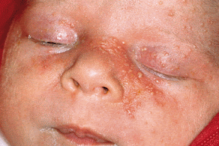
Introduction to Pediatric Milia


Milia are benign, self-limited lesions that manifest as tiny white bumps on the forehead, nose, upper lip, and cheeks of the newborn.

Pathophysiology
Milia is observed as small multiple cysts ranging from 1-2 mm in diameter. Histologically, these cysts are multiple superficial inclusion cysts that involve the follicular infundibulum (upper part of the hair follicle). They contain keratin and surrounded by a dense lymphocytic infiltrate. No visible opening is present.

Age of Onset

Rash appears in neonates 1-2 days after birth. It can be delayed for days to weeks in neonates born prematurely.

Clinical Presentation
The milia lesions range from 1-2 mm in size and are papular. They are pearly opalescent lesions and mostly present on the face. These lesions are called Epstein pearls when present on the soft or hard palate.

Chickenpox In Children


Varicella, commonly known as chickenpox, is caused by the varicella-zoster virus. The disease is generally regarded as a mild, self-limiting viral illness with occasional complications. Varicella is common and highly contagious and affects nearly all susceptible children before adolescence.

Chickenpox is usually a benign disease in children, and almost all children recover uneventfully. However, varicella is not totally benign even today. A significant number of varicella cases are associated with complications, among the most serious of which are varicella pneumonia and encephalitis.

Pathophysiology
The causative organism, varicella-zoster virus, is a member of the human herpesvirus subfamily.

The virus enters through the respiratory system (conjunctival or upper respiratory mucosa) and colonizes the upper respiratory tract. Viral replication takes place in regional lymph nodes over the next 2-4 days; 4-6 days later, a primary viremia spreads the virus to reticuloendothelial cells in the spleen, liver, and elsewhere.

After a week, a secondary viremia disseminates the virus to the viscera and skin, eliciting the typical skin lesions.

This viremia also spreads the virus to respiratory sites and is responsible for the contagion of varicella before the appearance of the rash. Infection of the central nervous system (CNS) or liver also occurs at this time, as may encephalitis, hepatitis, or pneumonia.

The usual incubation period is 10-21 days. The patient is contagious from 1-2 days before the appearance of rash until the lesions crust over, usually 5-6 days after the rash first appears.

Management of Pneumonia in Children


Treatment decisions in children with pneumonia are dictated based on the likely etiology of the infectious organism and the age and clinical status of the patient.

After initiating therapy, the most important tasks are resolving the symptoms and clearing the infiltrate. With successful therapy, symptoms resolve much sooner that the infiltrate.

The Pediatric Infectious Diseases Society and the Infectious Diseases Society of America created evidence-based guidelines for the management of community-acquired pneumonia (CAP) in infants and children older than 3 months. These guidelines discuss site-of-care management, diagnosis, antimicrobial therapy, adjunctive surgical therapy, and prevention. While these guidelines do not represent the only approach to diagnosis and therapy, these recommendations may assist in decreasing morbidity and mortality rates in children with community acquired pneumonia.

Hospitalization
Pulse oximetry should be performed during the prehospital evaluation of children with suspected pneumonia, and supplemental oxygen should be administered, if necessary; however, many school-aged children do not require hospitalization and respond well to oral antibiotics. Usually, these patients are not toxic or hypoxic enough to require supplemental oxygen. Unless they are vomiting, they do not require intravenous fluids or antibiotics. A parapneumonic effusion that requires drainage usually dictates a hospital admission.

Introduction to Ear Pain In Children


Otalgia or an earache
is ear pain. 

Primary otalgia is ear pain that originates inside the ear. 
Referred otalgia is ear pain that originates from outside the ear.

Otalgia is not always associated with ear disease. It may be caused by several other conditions, such as impacted teeth, sinus disease, inflamed tonsils, infections in the nose and pharynx.

Although ear pain can be troublesome and cause a difficult, sleepless night for both parents and child, it is rarely a reason to rush into emergency or to urgently start antibiotics.

Causes
Ear pain can be caused by disease in the external, middle, or inner ear, but the three are indistinguishable in terms of the pain experienced.

External ear pain may be:
Mechanical: trauma, foreign bodies such as hairs, insects or cotton buds.
Infective (otitis externa): Staphylococcus, Pseudomonas, Candida, herpes zoster, or viral Myringitis.

Middle ear pain may be:
Mechanical: barotrauma (often iatrogenic), Eustachian tube obstruction leading to acute otitis media.
Inflammatory / infective: acute otitis media, mastoiditis .

It’s not unusual for an ear infection to develop in early childhood. Although they’re not contagious, ear infections can occur as side effects of contagious illnesses—colds, coughs, or eye ailments.

Clinical History

Pain is the number one symptom indicating an ear infection. A child may tell that his ear hurts. Or, if he can’t talk yet but seems cranky or is tugging at his ear, an ear infection is a definite possibility. A recent cold or sinus infection is another clue. He may or may not have a fever.

Saturday, September 23, 2017

Meningitis - How to Diagnose it


Meningitis
, an inflammation of the membranes that cover the brain and spinal cord, can be caused by infection with a bacterium or virus.

Children who have partially treated meningitis or develop it while on antibiotics have modified signs and symptoms, and the diagnosis is usually delayed.

Bacterial meningitis is a life-threatening illness that results from bacterial infection of the meninges. Beyond the neonatal period, the 3 most common organisms that cause acute bacterial meningitis are Streptococcus pneumoniae, Neisseria meningitidis,and Haemophilus influenzae type b (Hib). Since the routine use of Hib, conjugate pneumococcal, and conjugate meningococcal vaccines in the United States, the incidence of meningitis has dramatically decreased.

Examination

Upon arrival at the emergency department, the child’s temperature, blood pressure, respiratory rate, pulse, and oxygen in the blood may be checked. After quickly checking the child’s airway, breathing, and circulation, the doctor completely examines the child to look for a focal source of infection, to assess any alteration in mental status, and to determine the presence of meningitis. If meningitis is suspected, several tests and procedures are needed to determine the diagnosis.

Investigations

Lumbar Puncture
The most important laboratory study is examination of CSF. The lumbar puncture (LP) should include opening and closing pressure in the cooperative patient.
  • Cell count
  • Gram stain
  • Culture and sensitivity
  • Glucose
  • Protein and antigen
  • Acid-fast bacillus
  • Fungal stains

Patau syndrome – Trisomy 13


Patau syndrome
, also known as trisomy 13 and trisomy D, is a chromosomal abnormality, a syndrome in which a patient has an additional chromosome 13 due to a nondisjunction of chromosomes during meiosis.

Causes
Trisomy 13 occurs when extra DNA from chromosome 13 appears in some or all of the body’s cells.
  • Trisomy 13 — the presence of an extra (third) chromosome 13 in all of the cells.
  • Trisomy 13 mosaicism — the presence of an extra chromosome 13 in some of the cells.
  • Partial trisomy — the presence of a part of an extra chromosome 13 in the cells.
Incidence
  • Patau syndrome is the least common and most severe of the viable autosomal trisomies.
  • Trisomy 13 occurs in about 1 out of every 10,000 newborns.
  • Median survival is fewer than 3 days, with only one in 20 children surviving longer than 6 months.
  • The sex ratio at birth is slightly skewed toward females, presumably because of decreased survival among males.
Clinical Presentation
Newborns with Patau syndrome typically present in the neonatal period with low Apgar scores and may have the following conditions:
  • Cleft lip
  • Cleft palate
  • Polydactyly (postaxial)
  • Microcephaly
  • Rocker-bottom feet
  • Microphthalmia
  • Scalp defects (cutis aplasia)
  • Omphalocele
  • Hernias
  • Neural tube defects
  • Stillbirth and in utero fetal demise are common pregnancy outcomes
  • Cardiac defects occur in 80% of cases
  • Holoprosencephaly, in which the brain does not divide completely into halves, is often present and is generally signaled by the presence of midline facial defects. Facial defects include the following:
  • Hypotelorism
  • Microphthalmia
  • Anophthalmia
  • Absent or malformed nose or proboscis
  • Severe clefting of the lip and/or palate.

Introduction to Patent Ductus Arteriosus


Patent ductus arteriosus (PDA)
is a condition in which the ductus arteriosus does not close. (The word “patent” means open.)

The ductus arteriosus is a blood vessel that allows blood to go around the baby’s lungs before birth. Soon after the infant is born and the lungs fill with air, the ductus arteriosus is no longer needed. It usually closes in a couple of days after birth.

PDA leads to abnormal blood flow between the aorta and pulmonary artery, two major blood vessels that carry blood from the heart.

A small patent ductus arteriosus often doesn’t cause symptoms or problems and may never need treatment. Untreated, a large patent ductus arteriosus can cause too much poorly oxygenated blood to flow through the heart, weakening the heart muscle and causing heart failure and other complications.

In some cases, such as in transposition of great vessels (the pulmonary artery and the aorta), a PDA may need to remain open. In this cardiovascular condition, the PDA is the only way that oxygenated blood can mix with deoxygenated blood. In these cases, prostaglandins are used to keep the patent ductus arteriosus open.

Pathophysiology
Failure of ductus arteriosus contraction in preterm neonates has been suggested to be due to poor prostaglandin metabolism because of immature lungs. Furthermore, high reactivity to prostaglandin and reduced calcium sensitivity to oxygen in vascular smooth muscle cells contribute to contraction of the ductus. The absence of ductus arteriosus contraction in full-term neonates might be due to failed prostaglandin metabolism most likely caused by hypoxemia, asphyxia, or increased pulmonary blood flow, renal failure, and respiratory disorders.