Tuesday, July 18, 2017

Approach To A Child Presenting With Urinary Retention


Acute urinary retention is relatively infrequent in children. There are a variety of causes that are poorly defined in the literature, and they differ greatly from those seen most frequently in adults.

Urinary retention means an inability to void or unable to empty the bladder.

Taking the History

When a child presents with the complains of inability to void a detailed history is required in order to reach the diagnosis and then manage accordingly.

Ask:

1. If the patient is toilet trained?

2. Is patient constipated

3. Is there pain on urination or fever

4. Any history of trauma

5. Past history of UTI

6. Any history suggestive of sexual abuse

7. Any medications currently in use

Physical Examination
After a detailed history relevant physical examination is necessary that may include
  • General Physical Appearance: the child may appear ill looking and uncomfortable.
  • Abdomen: Tender , tense smooth suprapubic mass usually indicates a distended urinary bladder.
  • Genitalia: Phimosis ( if uncircumcised) , meatal stenosis and erythema of perpuse or glans may represent acute balanoposthitis or erythema may indicate sexual abuse.
  • Neurological examination: is needed to assess the sensation in the perineal area.

Asthma – Management in Emergency Room


A child with acute attack of asthma will usually present with difficulty breathing, cough, wheezes and cyanosis.

Initial Assessment
Assess the Heart Rate, Respiratory Rate, O2 saturation, Peak expiatory flow rate, Use of accessory muscles, Pulsus paradoxus ( more than 20 mmHg difference in systolic B.P for inspiratory versus expiratory phase ) , Dyspnea, Alertness, Colour.

Initial Management

1. Give O2 to keep saturation > 95%.

2. Administer inhaled B- agonists: Nebulized albuterol 0.05 to 0.015 mg/kg/dose every 20 minutes or continuously depending on the condition.

3. Other nebulized bronchodilators that can be used include ipratropium bromide 0.25 to 0.5 mg.

4. If the air movement is poor or the patient is unable to cooperate with a nebulizer give epinephrine 0.01 ml / kg SC . It can be given every 15 minutes upto 3 doses.

5. Starting Steroids: If there is no response after one nebulized treatment or if the patient is steroid dependent or had a recent emergency depertment visit or an ICU care needed start prednisolone 2 mg/kg /day divided 6 hrly.

Blood Components and Their Uses in Pediatrics


Many blood products are available but they have never been safe as they can transmit diseases. For this reason children should only recieve blood products when othe conservative measures have failed.

Red Blood Cells ( RBCs )
Indicated in cases of severe anemia usually when Hb is < 7 gm%. or when there is acute , severe, traumatic blood loss.

Different types include:
  • Whole Blood ( rarely used )
  • Packed RBCs = whole blood less 70% of plasma , most commonly used
  • Leukocyte poor RBCs= for pateints with history of febrile reaction to blood products or who will recieve many transfusions
  • Washed RBCs = to prevent host-versus graft disease in Ig A deficient recipients

Respiratory Distress Syndrome Of Newborns - A Brief Discussion


Introduction
Respiratory Distress Syndrome of the newborns (RDS) also known as hyaline membrane disease is a breathing disorder of premature newborns in which the air sacs (alveoli) in a newborn’s lungs do not remain open because the production of a substance that coats the alveoli (surfactant) is absent or insufficient. This disease is mainly confined to premature babies. Insufficient surfactant leads to alveolar collapse; re-inflation with each breath exhausts the baby, and respiratory failure follows. Hypoxia leads todecreased cardiac output, hypotension, acidosis and renal failure. It is the
major cause of death from prematurity.

Risk factors:

  • Preterm babies ( 91%risk if born at 23–25 weeks; 52% risk  if 30–31 weeks.)  
  • Maternal diabetes,
  • Male babies, 
  • 2nd twin, 
  • Caesarean deliveries.

Clinical Signs 
  • Respiratory distress shortly after birth (within first 4 hours)
  • tachypnoea ( respiratory rate >60/min),
  • grunting, 
  • nasal flaring, 
  • intercostal recession and 
  • cyanosis. 
Chest X ray shows diffuse granular patterns (ground glass appearance) ± air bronchograms.

Saturday, July 15, 2017

Heel Stick Capillary Blood sampling


Heel stick is a minimally invasive and easily accessible way of obtaining capillary blood samples for various laboratory tests, especially newborn screens and glucose levels.

Indications

For the collection of blood in neonates and infants.

Contraindications

Edematous skin puncture site
Children older than 1 year of age.

Materials need for the procedure

  • Sterile gauze
  • Alcohol wipe
  • Heel warmer
  • Automatic lancet device
  • Appropriate containers for blood sample
  • Adhesive bandage
  • Sterile gloves
Procedure

1. Warm the infant’s heal for approximately 5 minutes. With gloved hands , cleanse the area with alcohol wipe. Hold the foot firmly with the leg in a tucked – flexed position

2. Grasp infant’s heel in a moderately firm grip, with the fore finger at the arch of the foot and the thumb below the puncture site at the ankle.

Approach to a Child With Eye Discharge and Swelling


Introduction
Conjunctivitis is a common infection especially among children under five. Children with conjunctivitis must be kept home from school or day care until the discharge from their eyes has stopped. This will prevent the spread of infection to other children. The incidence of conjunctivitis decreases with age.
Conjunctivitis leads to:
  • Eye irritation and redness
  • Excessive tears in the eyes
  • A discharge with pus
  • Swelling of the eyelids
  • Photophobia
History Taking
Age
Age of the child is important in determining the etiology of conjuctivitis. In newborns typical cause is chemical, chalmyadial or bacterial. In older children bacteria , viruses and allergies are more likely causes.

Onset of Symptoms
Time of onset is especially important in the neonatal period. With chemical conjunctivitis inflammation begins a few hours after drops have been placed and lasts for 24 to 36 hrs. Gonococccol conjunctivitis develops between 2 and 5 days of life.

Characteristic of discharge

  • Viral conjunctivitis usually causes watery or serous discharge.
  • Bacteria causes purulent or mucopurulent discharges.
  • Allergies produce serous or mucoid discharge often very stringy.
Associated Symptoms
Viral conjunctivitis is often associated with upper respiratory symptoms or other systemic complains like pharyngitis . Preauricular adenopathy and rashes may also be present. With vesicles or corneal ulceration HSV must be rules out. Pain on movement of the eyes is never normal and orbital cellulitis should be ruled out.

Umbilical Vein Catheterization


Umbilical vein catheterization may be a life-saving procedure in neonates who require vascular access and resuscitation. The umbilical vein remains patent and viable for cannulation until approximately 1 week after birth. After proper placement of the umbilical line, intravenous fluids and medication may be administered to critically ill neonates.

Indications
  • Emergency administration of medications and fluids in the delivery room or neonatal intensive care unit.
  • Blood sampling.
Contraindications
  • Omphalitis
  • Necrotizing enterocolitis
Materials needed for the Procedure
3.5 Fr or 5 Fr umbillical catheter, Heparinized saline( 0.5-1 unit/ml ) , 3 way stop cock, syringes, 10% poviodone-iodine preparation, sutures, adhesive tape, sterile cutdown tray, sterile gloves, mask, protective eye wear.

Procedure

1. Put on mask, protective eye wear and sterile gloves. Use aseptic technique and observe standard precautions through out the procedure.

2.Flush a 3.5 Fr or 5fr radiopaque umbilical catheter with heparinized saline ( 0.5 – 1 unit/ml ) attached to a 3 way stopcock.

Transposition of the Great Arteries


Transposition of the Great Arteries
is one of the cyanotic congenital heart disorders in which the two main arteries leaving the heart are reversed (transposed).

Transposition of the great vessels is a cyanotic heart defect. This means there is decreased oxygen in the blood that is pumped from the heart to the rest of the body.

In normal hearts, blood that returns from the body goes through the right side of the heart and pulmonary artery to the lungs to get oxygen. The blood then comes back to the left side of the heart and travels out the aorta to the body.

In transposition of the great vessels, the blood goes to the lungs, picks up oxygen, returns to the heart, and then flows right back to the lungs without ever going to the body. Blood from the body returns to the heart and goes back to the body without ever picking up oxygen in the lungs.

Etiology
Etiology for transposition of the great arteries is unknown and is presumed to be multifactorial. Preexisting diabetes mellitus of an expectant mother is a risk factor that has been described for the fetus having TGV.

Clinical Presentation
Infants with transposition of the great arteries (TGA) are usually born at term, with cyanosis apparent within hours of birth.

The clinical course and manifestations depend on the extent of intercirculatory mixing and the presence of associated anatomic lesions.

The common findings include:
  • Blueness of the skin
  • Clubbing of the fingers or toes
  • Poor feeding
  • Shortness of breath.

Hypothermia in Pediatric Practice

Hypothermia is defined as a core temperature of 35 C ( 95 F ) or less. It occurs when the body is no longer able to sustain normal body temperature. Onset of hypothermia depends on the imbalance between increased heat loss and decreased heat production.

Immediate Assessment and Questions
  • What are the vital signs? Is there a Pulse?
  • Is the patient breathing?
  • What is the clinical setting? Very young children are susceptible to hypothermia as a result of environmental exposure.
  • Any history of other medical problems like hypoglycemia, hypopituitarisim and hypoadrenalisim may present with hypothermia.
  • Any possible source of infection. Septic patients may present with hypothermia.
Physical Examination
  • Vital signs
  • Measure core body temperature , pulse and B.P
  • Volume status
  • Volume status should be continuously monitored. Observe for signs of trauma or any continuing blood loss.
Neurological exam
Check mental status and perform a complete neurological exam if the patient is not comatose.

Workup
  • CBC: To help rule out conditions such as sepsis and to determine Hb concentration and oxygen carrying capacity.
  • Basic Metabolic panel: Provides clues to volume status, hypoglycemia and electrolyte disturbance.
  • Urinalysis
  • ABGs : Provides acid-base status a nd oxygenation status.
  • Chest X-ray: To look for pulmonary edema or aspiration
Physiology of Hypothermia in childrenChildren have a higher surface area to mass ratio than adults and therefore cool faster and become hypothermic at an increased rate. Environmental factors like cold, wind and inadequate clothing can increase heat loss. Heat production can be affected by such factors as age, trauma and medical illness.

Stages of Hypothermia

  1. Mild – Core temperature 32 to 35ºC (90 to 95ºF)
  2. Moderate – Core temperature 28 to 32ºC (82 to 90ºF)
  3. Severe – Core temperature below 28ºC (82ºF)

Differential Diagnosis of Diarrhea in Children


Children may present with diarrhea which is defined as the passage of loose stools. It is a very common complaint and a list of differential diagnosis should be kept in mind while dealing with a child presenting with this problem.

1. Infection

Viral: Usually resolves in a few days and can be treated symptomatically. Rotavirus, adenovirus and Norwalk virus are more common.

Bacterial: Shigella, salmonella, Campylobacter, E coli , Vibrio, Clostridium species are commonly involved. Spectrum of illness may range from asymptomatic to life threatening. Staph aureus and C perfringens are often associated with food poisoning. Enterohemorrhagic E coli causes bloody diarrhea and may be associated with hemolytic uremic syndrome.

Parasitic: Giardia is often contracted by drinking contaminated water and can cause abdominal distension, diarrhea and at times failure to thrive. Giardia is easily passed through fecal oral route and is common in day care centers. Entamoeba histolytica produces severe coilitis.

2.Postinfectious State
Several viruses can cause severe enteritis resulting in prolonged intestinal mucosal damage, acquired carbohydrate intolerance and malabsorption in infants.

Failure to Thrive – Differential Diagnosis


A child may present with failure to thrive and not gaining appropriate weight for his age. There are a number of different factors responsible for it and a doctor should consider the following differentials in his mind.

Inadequate Intake
May result from a swallowing problem, formula mixing error, lack of access to formula or other foods due to limited finances, inappropriate substitution of other liquids ( eg juices ) for formula or neglect.

Excessive losses due to Diarrhea

Consider infectious agents ( bacterial, giardia ), HIV, cystic fibrosis or inflammation due to food allergies.

Increased Needs
It may be due to hyper metabolic states or disease that cause increased work of breathing. Consider cystic fibrosis, hyperthyroidism and cardiac disease.

Nausea Treatment in Cancer Patients


A child diagnosed with any malignancy may present with nausea and vomiting and a proper care and treatment is necessary.

Etiology
Usual cause is side effect of chemotherapy treatment. Also suspect opiate therapy, GI and CNS radiotherapy, obstructive abdominal mass, CNS mass, certain antibiotics or hypercalcemia.

Presentation

Acute: Emesis within 24 hrs of starting chemotherapy occurs in one third of patients despite treatment.

Delayed: Emesis occuring 24 hrs after chemotherapy , increased risk in females, prior acute emesis and with certain agents e.g ciplatin.

Anticipator
y: Emesis that occurs prior to chemotherapyadministration.

Treatment


Hydration plus one or more antinausea medications are needed.

1. Serotonin (5-HT3 ) antagonists
Ondansetron, dolasetron, granisetron. Usually a first line therapy. Patients may respond preferentially to one of these agents. Beware of QT prolongation, widening of QRS .

2. Histamin 1 Antagonist

Diphenhydramine, also cyproheptadine with anticholinergic side effect of appetite stimulation.

3. Steroids

Dexamethasone especially helpful in aptients with brain tumors. Synergy of unknown mechanism with 5-HT 3 antagonists.

Friday, July 14, 2017

Umbilical Granuloma - Clinical Features & Management

Introduction:
An umbilical granuloma is granulation tissue with incomplete epithelialization that persists following cord separation.
It is the most common cause of an umbilical mass in neonates.

Clinical Features: 

       Umbilical Granuloma. Newborn infant with umbilical granuloma visible in umbilicus

  • Parents will describe a persistent discharge from the umbilicus after the cord has dried and separated. It appears soft, pink, wet, and friable. 
  • Infants with an umbilical granuloma do not have localized swelling, redness, warmth, tenderness, or fever. 

Differential Diagnosis: 

  • An umbilical polyp is a rare anomaly resulting from the persistence of the omphalomesenteric duct or the urachus and may have a similar appearance. A polyp is usually firm with a mucoid secretion. 
  • The differential diagnosis also includes omphalitis, an infection of the umbilicus and surrounding structures, which should be considered in ill appearing neonates.

Wednesday, July 12, 2017

Knee Arthrocentesis


Knee Arthrocentesis or joint aspiration
is a procedure whereby a sterile needle and syringe are used to drain joint fluid from the knee joint.

Indications

1. Diagnostic
  • Removal of fluid for culture if suspicious of septic joint.
  • Removal of fluid if suspicious of hemarthrosis or traumatic joint injury.
2. Therapeutic
  • Relief of discomfort caused by intra articular pressure and distension of joint capsule and ligaments by accumulating fluid.
  • Drainage of pus to decrease damaging effects of bacteria, WBCs and inflammatory response on joint space.
  • Injection of medications such as glucocorticoids ( one of the modalities of treatment in non infectious inflammatory arthritis such as juvenile rheumatoid arthritis )
Contraindications
  • Hemophilia
  • Overlying skin infection or cellulitis
  • Bacteremia
  • Anatomic inaccessibility

Management Of Anaphylaxis In Emergency Room


Definition of Anaphylaxis

Anaphylaxis is the clinical syndrome of immediate hypersensitivity. It is characterized by cardiovascular collapse, respiratory compromise and cutaneous and GI symptoms ( e.g urticaria, emesis )

Initial Management


1. ABC’s :
Establish airway if necessary. Assess breathing. Supply with 100% oxygen with respiratory support as needed. Assess circulation and establish IV access. Place patient on cardiac monitor.

2. Epinephrine: Give epinephrine , 0.01 ml/kg (1:1000) intramuscular, maximum dose 0.5 ml. Repeat every 15 min as needed. The site of choice is lateral aspect of thigh due to its vascularity.

3. Albuterol: Give nebulized albuterol, 0.05 to 0.15 mg/kg in 3 ml normal saline solution ( quick estimate 2.5 mg for < 30kg and 5 mg for > 30kg ) every 15 min as needed.

4. Histamine 1- receptor antagonist: such as diphenhydramine, 1-2 mg/kg through IV.IM or oral route. Also consider a histamine -2 receptor antagonist.

5.Corticosteroids: helps prevent the late phase of allergic response. Administer methlyprednisolone in a 2 mg/kg IV bolus, then 2mg/kg per day IV or IM divided every 6 hrs or prednisone 2 mg/kg PO in a bolus once daily. Observe for 6 to 24 hrs for late phase symptoms depending on clinical condition and stability.

A Child Presenting With Cyanosis – Differential Diagnosis


Cyanosis
refers to the bluish skin color attributable most often to the presence of desaturated hemoglobin ( 5g/dl ). Primary etiologies include respiratory, cardiac, circulatory and nervous system disorders, as well as abnormal hemoglobin.

Respiratory Diseases

1. Lung disease

  • Newborn: Lung hypoplasia , Diaphragmatic hernia, respiratory distress syndrome, transient tachypnea of the newborn, Bronchopulmonary dysplasia, Pulmonary interstitial emphysema, Congenital adematoid malformation, Meconium aspiration.
  • Infectious: Pneumonia, Pneumonitis, Bronchiolitis.
  • Asthma
  • Cystic fibrosis
  • Infiltrative disease: Pulmonary hemosiderosis, sarcoidosis
2. Airway abnormalities or Obstruction
  • Congenital: choanal atresia, macroglossia, micrognathia, laryngeal web, tracheal stenosis, vascular ring, tracheoesophageal fistula.
  • Infectious: Acute epiglottitis, croup, retropharyngeal abscess, laryngospasm.
  • Traumatic: Vocal cord injury, pneumothorax, pneumomediastinum
  • Others: lymphoma, cystic hygroma, goiter, laryngeal hemangioma or neoplasm, foreign body, obesity.
3. Trauma
  • Pneumothorax, pneumomediastinum, vocal cord injury.
Pulmonary Vascular Diseases

1. Primary pulmonary hypertension
  • Cyanosis requires a right to left shunt at the atrial or ductal level unless lung disease is present.
2. Pulmonary arteriovenous malformation
  • Idiopathic or associated with congenital heart, hepatic or portal disease.

Workup In A Child Presenting with Neck Swelling And Masses


A child may present with a complaint of swelling or masses in the neck. In such a case apart from a complete history from the parent and a detailed physical examination some of the laboratory workup is important to reach a proper diagnosis and then appropriate management.

Laboratory Data


CBC with Differentials: May show an elevated WBC count in infectious processes, including cervical adenitis, retropharyngeal abscess and peritonsillar abscess. The test should also be performed if malignancy is suspected.

Blood Chemistries:
including renal and hepatic function tests and urinalysis.

Blood culture: can help guide to proper antibiotics if positive.

Gram stain, areobic and anareobic cultures:
can be obtained through needle aspiration or incision and drainage; may reveal causative agent in the diagnosis of acute cervical lymphadenitis or abscess.

Purified protein derivative ( PPD ) skin testing: it is recommended for children with subacute or chronic cervical lymphadenitis to rule out Mycobacterium tuberculosis especially if risk factors are present or there is poor response to initial treatment.

Other laboratory tests:
depending on the history and physical examination specific tests may be considered for example Bartonella henselae for cat scratch disease and Monospot test for EBV.

Fever Of Unknown Origin – Differential Diagnosis


Definition:
Fever of unknown origin is defined as a fever in which patient has a temperature of more than 101F (38.3 C ) on most of the days for at least 3 weeks duration of a single illness and the diagnosis is unclear despite 1 week of intense inpatient investigations.

The list of potential etiologies of FUO is vast but following can be considered:

1. Infection
In almost all reviews of FUO in pediatric patients , infection is the largest category. It is important to recognize uncommon manifestations of common disorders. About half of the localized infections involve the respiratory tract and a careful history and x-ray can confirm the diagnosis. Other locations that are sources of prolonged fever include urinary tract, bone and CNS. Look for clues to more generalized infections like epstein-barr virus , enteric fever, cat-scratch disease etc in which there may be evidence of multiple organ involvement.

2. Collagen or Connective tissue Disease
Juvenile rheumatoid arthritis may present with a long duration of fever before a diagnosis is established. Additional causes include Kawasaki disease, SLE, rheumatic fever and other vasculitic syndromes. Some of these diseases may produce additional physical findings but some patients have only atypical presentations with only a few manifestations of the disorder.

3. Neoplasia
Most common in this group are lymphoreticular malignancies ( e.g lymphoma. leukemia ). If there are any joint symptoms , there may at times be confused with JRA. Neuroblastomas and occasionally other sarcomas may also sometimes present with fever as the only major symptom.

Introduction to Reactive Arthritis In Pediatric Practice


Reactive arthritis
although not much commonly seen in pediatric practice is defined as a group of inflammatory arthritis which follow bacterial or viral infection particularly involving respiratory, gastrointestinal and genitourinary tracts.

Mode of Onset

Infection typically precedes development of arthritis by 1 to 4 weeks, with approximately 80% of cases being preceded by gastroenteritis.

Some Precipitating Organisms

Mycoplasma, Chlamydia, yersinia, Salmonella, Shigella, Campylobacter, Epstein barr virus, Parvovirus B19,and enteroviruses.

Clinical presentation
Patient presents with symptoms like fever, weight loss and fatigue as well as dermatological and opthalmological findings. For example Reiter syndrome is a reactive arthritis in the presence of conjunctivitis and urethritis.

Diagnostic Workup For A Child Presenting With Lethargy


A child may present with lethargy, altered mental status or drowsiness. Lethargy can be a manifestation of a number of clinical conditions , ranging from toxic and metabolic conditions to seizures. The differential diagnosis is broad and a proper workup is needed apart from a detailed history and physical examination.

Laboratory Data

Basic Metabolic Panel
Electrolyte abnormalities ( hypo/hypernatremia, hypo/hyperkalemia, hypo/hypercalcemia ) and hypo/hyperglycemia can present with alteration of mental status. Matabolic acidosis may point to an underlying metabolic disorder or intoxication. Respiratory acidosis can be seen in intoxications with alcohol, benzodiazepines and barbiturates.

Renal and Hepatic Profile

Can indicate acute or chronic end organ dysfunction.

CBC with differentials
Elevated WBC count may indicate acute infection.

Toxicology Screening and Drug levels
Based on patient’s history, order blood levels of prescription medications, ingested medications or substances of abuse.

ABGs
Obtain if hypoxia or acid base problems are suggested by history or physical examination.

Saturday, July 8, 2017

Epistaxis In A Child – Important Points in History


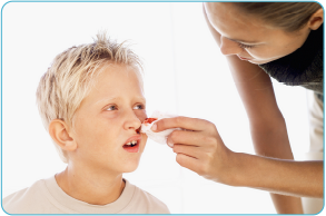
Epistaxis which is defined as bleeding from the nose is a common complain seen in children but usually the cause is not serious. A mother or the witness may be alarmed by the symptom and a child presenting with epistaxis needs to be asked following important questions in the history :

1. How old is the patient ?
Nose bleeds are unusual in very young children. In adolescent boys , juvenile nasopharyngeal angiofibroma is a rare benign, highly vascular neoplasm of the nasopharynx that can cause significant bleeding.

2. Was there any trauma to the nose ?
Allergic rhinitis can cause itching and subsequent rubbing of the nose , traumatizing the mucosa. Prior trauma from a direct blow on the nose may cause septal injury and bleeding. A dry environment causes crusting and young children may be prone to picking the nose , irritating septal mucosa. Unilateral foul or purulent discharge suggests a foreign body. In hospitalized patient, indwelling nasal tubes or suction trauma may injure mucosa and promote bleeding.

3. What medications has the patient taken ?
Use of NSAIDs promotes bleeding, particularly in children with undiagnosed coagulopathy. Antihistamines may thicken secretions and causes drying and cracking of mucosa. Nasal sprays , particularly those not in an aqueous vehicle or that rely on propellants can traumatize mucosa. patients should be instructed on proper way to spray nasal medications, directing stream away from septum. Herbal or alternative medicine may be associated with bleeding.

4. Is there a history of recent surgeries ?
Procedures requiring nasal intubation can injure mucosa. Adenoidectomy, sinus surgery and repair of nasal fracture among other procedures all carry a small risk of postoperative bleeding.

Introduction To Different Skin Erythemas In Children

The word Erythema is derived from the greek word erythrosthat means redness of the skin. It may occur with skin injury, infection or inflammation. Erythema disappears with finger pressure (blanching), while purpura or bleeding in the skin and pigmentation do not. There are few conditions that needs a short discription here:

1. Erythema Multiforme
Acute self limiting onset of symmetrical fixed red papules , some of which form target lesions. May blister. Can show koebner phenomenon. May involve lips, buccal mucosa, and tongue.


Causes: the causes of erythema multiforme includes:
  • Infections like herpes simplex virus, mycoplasma, EBV, Chylamydiae
  • Drugs like sulphonamides, penicillin
  • Collagen diseases like SLE, polyarteritis nodosa
  • Underlying malignancy
2. Steven- Johnson syndrome


Causes are same as for erythema multiforme.
Other features include
  • Severe erosions of at least two mucosal surfaces .
  • Prodromal respiratory illness.
  • Extensive necrosis of lips and mouth.
  • Purulent conjunctivitis
  • Variable skin involvement- red macules, bullae, skin necrosis and denudation.
3. Erythema nodosum
Nodular, erythematous eruption on extensor aspects of legs , less commonly on thighs and forearms. Regresses to bruises. May last for 3 to 6 weeks.

Causes: may include
  • Infections ; organisms mostly involved are streptococcus, salmonella, yersinea, tuberculosis, chlamydia, EBV etc
  • Gut disorders like ulcerative colitis and crohn’s disease
  • Malignancy like leukemias and lymphomas
  • Drugs e.g sulphonamides

Causes Of Hypotonia In The Newborn


A newborn child may be hypotonic and a number of different causes are associated with this condition. The following are the important causes of hypotonia in a newborn child.

Central Causes
  • Neonatal encephalopathy
  • Intracranial haemorrhage
  • Infection – generalized sepsis, meningitis, encephalitis.
  • Chromosomal abnormalities – trisomy 21, 18 or 13.
  • Structural brain abnormalities – neuronal migration disorders etc
  • Metabolic diseases – amino and organic acidaemias, urea cycle defects, galactosemia, non-ketotic hyperglycinaemia, mitochondrial disorders.
  • Drugs – opiates, barbiturates benzodiazepienes etc
  • Prader-Willi syndrome
  • Hypothyroidism
  • Early Kernicterus
Spinal Cord Lesions
  • Trauma to the cervical spinal cord during delivery- usually involves traction and rotation with forceps.
  • Tumors, cysts and vascular malformation of spinal cord.

Examination of the Neonate - A Quick Review


The aim is to screen for abnormality, and to see if the mother has any questions or difficulties.
The following is a recommended routine before the baby leaves hospital—or during the 1st week of life for home deliveries.

Before the examination find out :

  • If the birth weight was normal?
  • Was the birth and pregnancy normal? 
  • Is mother Rh–ve? 

For Examination 

  • Find a quiet, warm, well-lit room. 
  • Enlist the mother’s help. 
  • Explain your aims. 
  • Does she look angry or depressed? 
  • Listen if she talks. 

Examine systematically, from head-to-toe. 

  • Wash your hands meticulously.
  • Note observations (eg T°; rectal is more reliable than tympanic). 

Head:

  • Circumference (50th centile=35cm, ), 
  • shape (odd shapes from a difficult labour soon resolve), 
  • fontanelles (tense if crying or intracranial pressure increased; sunken if dehydrated). 

Eyes:

  • Red reflex (absent in cataract & retinoblastoma);
  • corneal opacities; 
  • conjunctivitis. 

Ears:

  • Shape; 
  • position. Are they low set (ie below eyes)? 

Nose: 

  • The tip of the nose, when pressed, shows jaundice in white babies.
  • Breathing out of the nose (shut the mouth) tests for choanal atresia
  • Ensure oto-acoustic screening is done 

Complexion: 

  • Cyanosed, pale, jaundiced, or ruddy (polycythaemia)?

Friday, July 7, 2017

Arterial Puncture In Children


Arterial puncture is done to obtain blood from an artery. The procedure is summarized as follows:

Indications
  • Blood gas determination
  • Arterial blood sampling to determine blood chemistries.
Contraindications
  • Poor collateral circulation.
  • Skin infection near planned puncture site.
  • Bleeding disorders, including thrombocytopenia, systemic fibrinolytic states and thrombolytic therapy.
Materials Needed for the procedure
Blood gas sampling kit,or 3-5 ml syringe, 1 ml heparin (1000 units/ml), alcohol or povidone-iodine swabs, and a cup of ice, 23-25 gauge needle (20-22 gauge for femoral artery), sterile gloves, sterile dressing, adhesive tape.

Procedure

1. Use a heparinized syringe for blood gas and a non heparinized syrine for chemistry determinations. Obtain a blood gas kit.

2. The radial artery is the most frequently used site for arterial blood gas sampling.

3. It may be helpful to secure the dorsum of the wrist and place gauze behind the wrist to hyperextend the joint. Elevating the wrist will often bring the radial and brachial arteries closer to the surface.

4. Prepare the area with either povidone-iodine or an alcohol swab. Hold the syringe like a pencil with the needle bevel up and enter the skin at a 60 to 90 degree angle. Maintain slight negative pressure on the syringe.

5. Aspirate very slowly. A good arterial blood sample requires only minimal back pressure. If the vessel cannot be located , redirect the needle without taking it out
of the skin.

6. Obtain about 2 -3 ml of blood.

Introduction To Mental Retardation


Definition

Mental retardation is defined as significantly subnormal intellectual functioning for a child’s developmental stage, with problems in self care, home living, communication and social interaction.

It is defined statistically as cognitive performance that is 2SD below the mean of the general population as measures on a standard intelligence testing.

Levels Of Mental Retardation

Mild: Have a Stanford-Binet IQ score of 67 -52 and a WISC-III IQ score of 70 – 55 and are educable.

Moderate: Have a Stanford-Binet IQ score of 51 -36 and a WISC-III IQ score of 54 – 40 and are trainable.

Severe: Have a Stanford-Binet IQ score of 35 -20 and a WISC-III IQ score of 39 – 25 .

Profound
: Have a Stanford-Binet IQ score of <20 and a WISC-III IQ score of <24.

Caution must be exercised in interpretation , however because these categories do not reflect the actual functioning level of the tested individual. Children who perform in the severe or profound ranges of MR also are capable of responding to some educational intervention.

Etiology

The etiology of the CNS insult resulting in MR may involve genetic disorders, teratogenic influences, perinatal insults, acquired childhood diseases and environmental and social factors. Mild MR correlates with socioeconomic status although profound MR does not.

A Child Presenting With Diarrhea- Important Questions In History


Diarrhea is a common presenting complaint in pediatric practice and following questions are important to be asked from the parent as these help in determining the condition of the child and the cause of diarrhea.

Immediate Questions

1. How old is patient?
In a young infant the intestinal mucosa tends to be more permeable to water than in an older child or adult. This can result in greater net fluid and electrolyte losses. Diarrhea in the first few months of life requires more immediate attention.

2.What are the vital signs?
Tachycardia suggests volume depletion. Hypotension suggests hypovolemic or septic shock. fever implies an infectious etiology. Diarrhea with associated tachycardia, hypotension or fever should be evaluated immediately.

3. Is diarrhea grossly bloody?
Bloody diarrhea is caused by invasive bacterial infections, ischemic bowel or infarction, allergic phenomenon or inflammatory bowel disease. It requires more active and immediate intervention.

4. Is this acute or chronic problem?
Acute diarrhea: Usually a self limited disease, which can often be treated symptomatically. The most common cause is infection. Other common causes include drugs e.g antibiotics as well as excessive intake of high carbohydrate fluids or non absorbable fillers (e.g sorbitol).

Chronic diarrhea: defined as diarrhea that lasts longer than 4 weeks. Common causes include chronic non specific diarrhea (toddler’s diarrhea), lactose intolerance, milk-protein allergy, encopresis, irritable bowel syndrome, various infections, drugs and IBD. It can also present as an acute exacerbation.

5. Are there risk factors that suggest a specific cause?
Risk factors include day care, winter season, ill contacts, drugs, travel, animal exposure, constipation, excessive juice intake, poorly perpared or stored poultry or salads, untreated water sources, prior abdominal surgery, immunodeficiency and prematurity.

List Of Anatomic And Mucociliary Defects That Result In Recurrent Or Opportunistic Infections


The major components of host defense include an anatomic barrier, innate immunity and adaptive immunity. Integrity of the anatomic-mucociliary barrier at the interface between the body and its environment is essential for protection against infection. 

Here is a list of anatomic and mucociliary defects that can result in recurrent or opportunistic infections.

Anatomic defects in upper airways
  • Aspiration syndromes ( gastroesophageal reflux, ineffective cough, foreign body )
  • Cleft palate, eustachian tube dysfunction
  • Adenoidal hypertrophy
  • Nasal polyps
  • Obstruction of paranasal sinus discharge
  • Post-traumatic or congenital sinus tracts ( CSF rhinorrhea )
Anatomic defects in the tracheo-bronchial tree
  • Tracheo-esophageal fistula, bronchobiliary fistula
  • Pulmonary sequestration, bronchogenic cysts, vascular ring
  • Tumor, foreign body or enlarged nodes
Physiologic defects in upper and lower airways
  • Primary ciliary dyskinesia syndrome
  • Cystic fibrosis, Bronchopulmonary dysplasia
  • Bronchiectasis
  • Allergic diseases (allergic rhinitis, asthma)
  • Chronic cigarette smoke exposure

Initial Laboratory Evaluation Of Respiratory Distress In Newborn


A newborn baby may present to the pediatric nursery with respiratory distress that is manifested by tachypnea, intercostal retractions, reduced air exchange, cyanosis, expiratory grunting and nasal flaring. These are all non specific responses to an underlying serious illness. The following tests are needed in the initial evaluation:

1. Chest Radiograph:

To determine reticular granular pattern of Respiratory distress syndrome; to determine presence of pneumothorax, cardiomegaly and any life threatening congenital anomalies.

2. Arterial Blood Gases:
To determine the severity of respiratory compromise, hypoxemia and hypercapnia and type of acidosis; the severity helps in the treatment strategy.

3. Complete Blood Count:
Hemoglobin and hematocrit to determine anemia and polycythemia; white blood cell count to determine neutropenia and underlying sepsis; platelet count and blood smear to rule out DIC.

4. Blood Culture:
To recover the potential pathogen and also helpful to start the specific treatment against the involved organism.

5. Blood Glucose:
To determine presence of hypoglycemia, which may produce or occur simultaneously with respiratory distress and if present should be treated immediately. Also sometimes patient has stress hyperglycemia .

Differential Diagnosis Of Stridor In Children


Definition of Stridor

Stridor is a harsh, high pitched respiratory sound produced by turbulent airflow that is usually inspiratory but may be biphasic; it is a sign of upper air way obstruction.

Stridor is commonly seen in sick children and is due to a number of different cause. A list of differential diagnosis of stridor is given here:

Infectious Causes

  • Acute laryngotracheobronchitis
  • Epiglottitis
  • Pharyngitis
  • Parapharyngeal abscess
  • Bacterial tracheitis
  • Laryngopharyngeal diptheria
  • Laryngeal papillomatosis
  • Extrinsic inflammatory mass compressing the trachea (e.g tuberculosis)
Non Infectious Conditions
  • Foreign body aspiration
  • Angioneurotic edema
  • Spasmodic croup
  • Ingestion of caustic or hot fluid
  • Trauma, smoke inhalation
  • Laryngomalacia
  • Congenital subglottic stenosis
  • Extrinsic mass compressing the trachea ( cystic hygroma, hemangioma, vascular malformation)
  • Hypocalcemia
  • Vocal cord paralysis

Paracentesis In Children


Parecentesis
is defined as a procedure in which a needle or a catheter is inserted into the peritoneal cavity through the abdominal wall in order to remove the ascitic fluid. It may be for diagnostic or therapeutic reasons.

Indications

Diagnostic:
  • Evaluation of new onset of ascites.
  • Exclusion of Peritonitis.
  • Assessment of clinical deterioration in known cirrhotic patients.
Therapeutic:
  • Decompression of tense ascites in symptomatic patients ( dyspnea, abdominal discomfort, early satiety )
Contraindications
Almost all are relative contraindications.
  • Coagulopathy: consider platelet transfusion for platelet count <50,000 and FFP for INR> 1.5
  • Uncooperative patient: consider sedation
  • Multiple surgical operations: consider surgical consultation.
Materials needed for the procedure
  • Minor procedure tray,
  • 18-22 gauge 1.5 inch catheter over needle assembly,
  • Syring (10-30 ml for infants and small children; 20-60 ml for older children and adolescents),
  • Sterile specimen containers,
  • Blood culture bottles,
  • Povidone-iodine solution,
  • Pressure dressing,
  • Sterile gloves

Introduction To Bartter Syndrome


Bartter syndrome
is caused by an inborn autosomal recessive defect, in the Na-K-2Cl co transporter in the thick ascending limb of the loop of Henle, leading to NaCl and water wasting.

Clinical Symptoms
The patient presents with polyuria, polydipsia, episodes of dehydration, flattering growth and constipation. sometimes there may be maternal polyhydramnios with an affected fetus.

Pathophysiology
Since there is NaCl and water wasting the resultant ECF volume contraction causes secondary renin secretion and raised aldosterone levels, with avid Na and water reabsorption in the distal tubule and reciprocal K and H secretion into the urine. Imprtant to note is that blod pressure is normal. There is also increases renal prostaglandin E2 secretion.

The above changes produce the characteristic biochemical disturbance of hypochloraemic hpokalaemic alkalosis.

Diagnosis
Crucial to the diagnosis is the finding of inappropriately high levels of urinary Cl and Na- usually more than 20mmol/L ; urine Ca is normal or high.

A Brief Discussion Of CNS Tumors In Children


Brain Tumors in children tend to be located in the posterior fossa, in the midline, have greater differentiation and have slightly better survival figures than their counterparts in adults.

The anatomical grouping together of brain tumors masks their diverse biological differences. As a general rule brain tumors do not metastasize out of the CNS.

Treatment options for CNS tumors remains controversial but usually involves surgery and/or radiotherapy.

Clinical Features

Brian tumors are notoriously difficult to diagnose because of thier varied and often non-specific presentations. The mean time from onset of symptoms to diagnosis is usually around 5 to 6 months.

The most common presenting symptom is vomiting and headache. Other features that may be seen are changes in personality and mood, deterioration of school performance, growth failure, weight loss and seizures. Some children may present with disturbance of speech and developmental delay.

A brief discussion of different CNS tumors in children is given here:

Astrocytoma
  • Most commonly occurring brain tumor.
  • Range from low grade (benign) tumors, usually in the cerebellum, to high grade (malignant) tumors, usually supratentorial and in the brain stem.
  • The glioblastoma multiforme tumor has a near fatal prognosis.

Legg-Calve-Perthes Disease


This is a non-inflammatory deformity of the femoral head caused by a vascular insult leading to osteonecrosis of the capital femoral epiphysis.

Incidence
  • It affects more boys than girls (4:1)
  • Usually presents between 4 and 8 years of age. ( can have a secondary peak between 10 and 12 years ).
  • Caucasians are affected more frequently than other races.
  • Occurs in approximately 5.5 of 100,000 children per year (and therefore a lifetime risk of developing the disease is about 1 per 1200 individuals)
Etiology
Although the cause is not clear it occurs in those with a delayed skeletal maturation and also there is an increased incidence with a positive family history or in children with a history of low birth weight.

Clinical Features

  • Pain (may present with referred pain in the knee).
  • Decreased range of motion.
  • Limping gait.
  • There are 4 clinical stages: Initial stage, fragmentation, healing and residual.

Neonatal Mastitis - Clinical features & Management


Neonatal mastitis is an infection of the breast tissue that occurs in full-term neonates with a peak incidence in the third week of life. Females are affected more often than males in a 2:1 distribution.

Clinical Signs and Symptoms: Clinically, it manifests as swelling, induration, erythema, warmth, and tenderness of the affected breast. The ipsilateral axillary lymph nodes may be swollen. Approximately two-thirds have palpable fluctuance. In some cases, purulent discharge may be expressed from the nipple. Fever may be present in 25% of affected patients. Other systemic symptoms (irritability, decreased appetite, and vomiting) are less common but indicate a more severe infection if present. Bacteremia is rare.

Pathology: Staphylococcus aureus is the most common pathogen, causing 75% to 85% of cases. Rarely, gram-negative organisms or group B or D Streptococcus are the cause. If treatment is delayed, mastitis may progress rapidly with involvement of subcutaneous tissues and subsequent toxicity. In the initial stages, neonatal mastitis may mimic mammary tissue hypertrophy owing to maternal passive hormonal stimulation. Minor trauma, cutaneous infections, and duct blockage may precede this infection.